The Musculoskeletal System
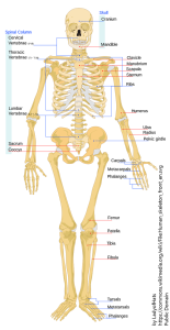
The Skeletal System is the framework of the human body and provides support, movement, protection, mineral homeostasis, hematopoiesis, and energy storage. Ligaments connect bones to other bones, and tendons connect bones to muscles. Movement is possible as muscles pull against the bone framework. Bones protect many vital internal organs (except the abdominal region). Bones also store calcium and phosphate which allows homeostatic mechanisms to maintain normal body levels of these critical minerals. Bone marrow is soft tissue that fills the cavities inside long bones. Red bone marrow is the location of hematopoiesis (red and white blood cell production) and yellow bone marrow is full of stored energy (in the form of triglycerides).
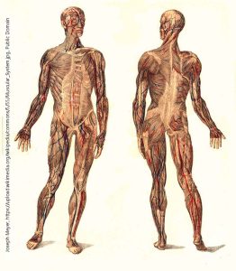
The Muscular System provides locomotion (movement), stabilization (posture), and thermoregulation. Various muscles and muscle groups give us either gross (large) or fine (small, precise) motor movement. There are three types of muscle tissue in the body:
- skeletal
- cardiac
- smooth
Just given their names, you probably already have an idea about where a couple of them are located.
Skeletal muscle is named such because of its connection to the skeleton. This relationship allows contractions of the muscles to move the body or maintain posture. Skeletal muscle is under voluntary control, meaning that contraction is controlled by conscious thought and intentional stimuli from the nervous system. Skeletal muscle also helps warm the body by burning energy to release heat (commonly called shivering). Skeletal muscle is striated (striped due to sarcomeres).
Cardiac muscle is found in just one place – the heart. It is not under voluntary control and is probably the hardest-working muscle in the body, contracting about 60-100 times per minute, every minute, every hour, every day of our lives. Cardiac muscle is striated and branches (intercalated discs). The purpose of cardiac muscle contraction is to squeeze the heart, pushing blood out of the chambers and into the arteries of the body. The branching arrangement of cells allows for this type of movement. Like skeletal muscle, the coordinated shortening of thousands of sarcomeres results in the contraction of the heart muscle. Cardiac muscle is unique in that it responds to signals that arise from specialized cells within the heart itself. These cells send electricals signals about every 0.8 seconds causing the heart to contract about 72 times per minute (called heart rate or pulse). When under stress, hormones, nervous system signals, or both can cause heart rate to increase.
Smooth muscle gets its name because it lacks the sarcomeres that give skeletal and cardiac muscle their striations. Smooth muscle contractions are typically slower to initiate, but last longer than other muscle types. Smooth muscle is located in the walls of hollow internal structures, such as blood vessels, airways, and the digestive system. It is also seen in other areas like the eyes and reproductive organs. Like cardiac muscle, it is not under voluntary control. Smooth muscle is responsible for vasodilation and vasoconstriction (increasing and decreasing blood vessel diameter), bronchodilation and bronchoconstriction (increasing and decreasing airway diameter), and peristalsis (wave movements propelling food through the gut, sperm through the ductus deferens and urethra, and ova through the uterine tubes).
Media Attributions
- Unit 5 figure 1 Skeletal System © LadyofHats, Mariana Ruiz Villarreal is licensed under a Public Domain license
- Unit 5 figure 2 Muscular System © Derivative of original by Bibliographisches Institut (copyright extinct) is licensed under a Public Domain license
