Talking About Molecules, Cells, and Tissues
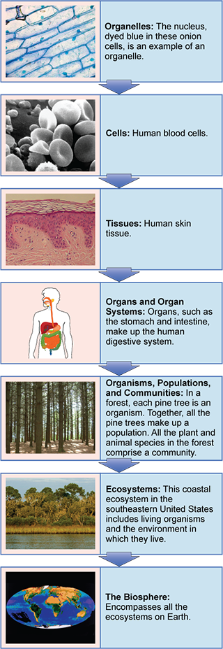
Living things are organized at different levels, from the atomic to the ecological. Human bodies are no different. A human body is made up of organ systems; organ systems are made up of tissues; tissues are made up of cells; cells are made up of molecules; and molecules are made up of atoms. (The word atom means indivisible or unable to be cut; before the 20th century, it was the smallest known unit of matter.)
We started our Medical English journey in Unit 1, looking at how Medical English is built. In Unit 2, we learned the Medical English used in clinics, and how the human body is described. Everyone who works in a clinical environment must use the same language to communicate effectively within and between health care teams. That’s why your professional education probably started with this course, Medical Terminology.
Part of becoming a clinician is understanding the workings of levels of organization smaller than the entire body: atoms, molecules, organelles, cells, and tissues. This Unit will familiarize you with the specialized language you need to excel in courses such as anatomy & physiology and pathophysiology.
Among the terms we’ll learn in this Unit are those that describe the parts of a cell. If you want to investigate this further, the web pages maintained by the National Cancer Institute are a good place to start. Here, we see some of the organelles and cell parts we’ll learn the names of:
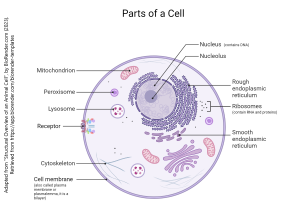
- the cell membrane, a bilayer made of phospholipids which surrounds the entire cell and keeps the insides in and the outsides out;
- membrane receptors, which allow the cell to interact with its environment, and react to external chemical signals like epinephrine, which are then turned by the receptor into electrical or chemical changes in the processes of the cell;
- the nucleus, which contains the genetic material, and the nucleolus, where the genetic material (DNA) is copied into RNA;
- the ribosome (not shown here), where RNA is turned into proteins, the workhorse molecule of the cell (ribosomes are often associated with the endoplasmic reticulum);
- mitochondria (singular mitochondrion), which makes the energy needed for proper cell function.
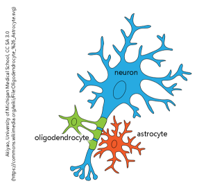
We’ll also look at different cell types which make up tissues. Some diseases involve changes in the number and distribution of cells in the body. For example, leukocytes are white blood cells, critical for the body’s immune defenses. If there are too many leukocytes of the wrong kind, then the disease leukemia results. Astrocytes (astroglia) are star-shaped cells which play an important role in maintaining brain function; if they become cancerous and their numbers increase uncontrollably, an astrocytoma may grow in the brain. Oligodendrocytes (oligodendroglia) have several arms that wrap parts of the nerve cell (neuron) in a protective layer. When they become cancerous, an oligodendroglioma may result.
Media Attributions
- Figure_01_02_15new 320 px wide © OpenStax is licensed under a CC BY (Attribution) license
- Unit 3 figure 2 Parts of a Cell © Jim Hutchins is licensed under a CC BY (Attribution) license
- Unit 3 figure 3 Cell Types © Akiyao from the University of Michigan Medical School adapted by Jim Hutchins is licensed under a CC BY-SA (Attribution ShareAlike) license
