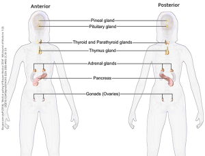
The Endocrine System
Only [Med Terms] in the Building Episode 7
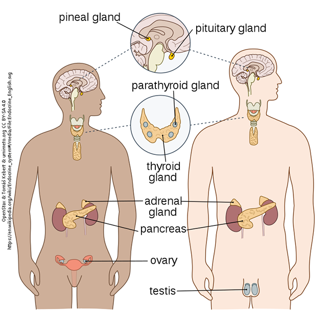
The Endocrine System, together with the nervous system, controls everything that occurs in the body’s internal environment. Some endocrine organs act independently, without nervous system control. Other times, the nervous system controls the endocrine gland. When the nervous system causes a gland to secrete, that gland is part of the endocrine system, and the secreted substance is a hormone.
The endocrine system is comprised of primary endocrine glands (hormone secretion is their only job) and secondary endocrine glands/tissues (secreting hormones is their second job).
| Hormone | Origin | Target | Action |
|---|---|---|---|
| adrenaline | epinephrine | adrenal medulla | cells in many organs | stimulates sympathetic nervous system (i.e. “fight-or-flight response” when the body is under stress |
| adrenocorticotropic hormone (ACTH) | anterior pituitary | adrenal cortex | stimulates adrenal cortex |
| antidiuretic hormone (ADH) | hypothalamus & posterior pituitary | kidneys | stimulates the kidneys to conserve water so that less urine is produced |
| calcitonin | a subset of thyroid cells | bones & kidneys | helps to regulate calcium levels in the blood |
| cortisol | adrenal cortex | cells in many organs | stimulates metabolism; inhibits inflammation; part of a class of hormones called glucocorticoids |
| estrogens | ovaries | uterine lining; many other cells | stimulate development of female sexual characteristics & helps the female body prepare for pregnancy; a class of hormones, the most common of which is 17-β-estradiol |
| glucagon | pancreas | primarily liver cells | stimulates the creation of glucose (gluconeogenesis) and breakdown of the storage form of glucose (glycogenolysis) |
| growth hormone | anterior pituitary | primarily bones and muscles | promotes growth |
| insulin | pancreas | most cells in the body | stimulates cells to take in glucose, which decreases blood sugar levels |
| oxytocin | hypothalamus & posterior pituitary | uterus, mammary glands, & brain cells | stimulates the uterus to contract during childbirth; stimulates the mammary glands to release milk; promotes bonding to another person |
| parathyroid hormone (PTH) | parathyroid glands | bones & kidneys | helps to regulate calcium levels in the blood |
| releasing hormones (many different kinds) | hypothalamus | anterior pituitary | stimulate the cells of the anterior pituitary to make and release hormones |
| testosterones | testes | many cells in the body | stimulate development of male sexual characteristics; stimulate development of sperm; a class of hormones, the most common of which is dihydrotestosterone |
| thyroid stimulating hormone (TSH) | anterior pituitary | thyroid gland | stimulates the thyroid gland to produce T4 and T3 |
| thyroxine (T4) & triiodothyronine (T3) | thyroid gland | cells throughout the body | stimulate metabolism |
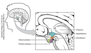
The hypothalamus is a part of the brain that produces hormones which either target or are stored in the pituitary gland. The pituitary gland is located inferior to the hypothalamus and has two parts, the anterior (adenohypophysis) and posterior (neurohypophysis). The hypothalamus controls the anterior pituitary (adenohypophysis) through a “private” blood system called the hypophyseal portal system. Releasing hormones are dropped into this private bloodstream and carried to the anterior pituitary, where they encounter cells that release hormones such as adrenocorticotropic hormone, growth hormone, and thyroid stimulating hormone.
The posterior pituitary (neurohypophysis) works differently. Neurons in the hypothalamus make the hormones antidiuretic hormone and oxytocin. They ship the hormone down their nerve cell axons which end in the posterior pituitary, where the release of hormones into the bloodstream occurs.
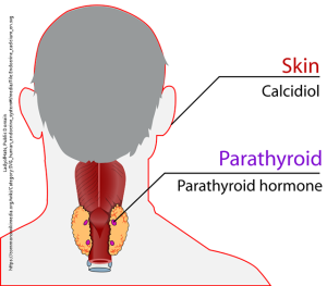
Another job of the endocrine system is regulating the amount of calcium carried in the blood. A small subset of thyroid gland cells releases calcitonin, and the nearby parathyroid glands release parathyroid hormone (PTH). The kidneys release calcitriol (vitamin D), a closely related compound, calcidiol, is made by the skin. Calcitonin moves calcium from the blood to the bones, lowering blood calcium levels; parathyroid hormone moves calcium from the bones to the blood, increasing blood calcium levels. Calcitriol (vitamin D) helps in the absorption of calcium in the intestines, which also increases blood calcium levels.
Hormones are also used to regulate sexual function. Genes on the X or Y chromosome will direct brain cells (through a complex, multi-step process) to begin producing hormones, which in turn promote the development of primary sexual characteristics (the gonads and external genitalia). Primary sexual characteristics are those a person is born with. After a pause of several years, another burst of hormones at puberty promotes the development of secondary sexual characteristics (for example, menstruation and breast development in the female). The beginning of menstruation is called menarche and the beginning of breast development is thelarche.
Media Attributions
- Unit 7 figure 1 endocrine system © OpenStax & Tomáš Kebert & umimeto.org is licensed under a CC BY-SA (Attribution ShareAlike) license
- Unit 7 figure 2 hypothalamus and pituitary © OpenStax College is licensed under a CC BY (Attribution) license
- Unit 7 figure 3 calcium homeostasis © LadyofHats is licensed under a Public Domain license
- Unit 7 figure 4 female endocrine system © BruceBlaus is licensed under a CC BY (Attribution) license
