The Nervous System
Only [Med Terms] in the Building Episode 6
The nervous system consists of the brain and spinal cord. The job of the nervous system is to control body processes; it does this by
- receiving information from the internal and external environment;
- processing that information in billions of neurons;
- sending that information to the brainstem and spinal cord, and from there to the muscles of the body.
It’s important to notice that the output of the nervous system is to move a muscle or change the secretion of a gland. That’s all it can do.
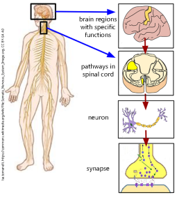
The nervous system is a triumph of miniturization as well as processing speed and efficiency. Imagine you’re driving and you see the brake lights of the car ahead of you. It’s time for a panic stop! Your visual system processes the lights and interprets what they mean. Visual association areas determine the correct action is to stomp on the brake as hard as possible. They send the command to an area called the premotor area, and the premotor area then writes up a work requisition and passes it to the primary motor cortex (the strip of brain shown in yellow in the top right of this diagram, also called the prefrontal gyrus). Nerve cables, called axons, carry signals through the spinal cord (second diagram from top right). The pathway (whose name is not important right now) is shown in yellow.
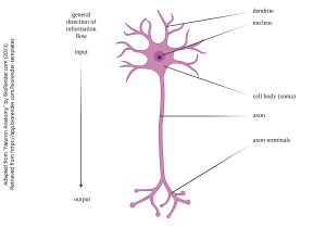
Eventually, the neurons that started off in the motor cortex end up in the anterior (ventral) horn of the spinal cord, where they send their signal to a motor neuron. This motor neuron picks up information at its dendrites. It has its cell body (soma) in the spinal cord, its axon in the nerves running to muscles, and ends in a synapse where it releases a chemical signal onto a muscle cell from its axon terminals. The chemical (acetylcholine) causes your thigh muscles to contract, and the brake is pressed, saving your life. That all happens in 100 milliseconds (0.1 second). Unfortunately, at 60 mph (27 m/sec) you have traveled about 1 car length while your brain has been processing the information. For this reason, neuroscientists don’t like it when you tailgate.
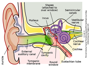
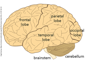
The auditory (hearing) system transforms sound waves which travel through the air into an electrical signal via the hair cells of the cochlea. Sound waves from your favorite music are transmitted for your enjoyment via the structures of the outer, middle, and inner ear to the temporal lobe of the brain.
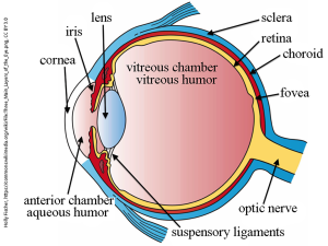
The eye is likely the most complicated of the sensory end-organs. It does an amazing job! It focuses a beam of photons onto the retina (a sheet of central nervous system cells at the back of the eye) and the retinal cells extract information (wavelength/color; edges; motion) from this beam of photons and send a report to the brain. The brain constructs a map of the visual world based on location and compares input from each eye. This helps us to more clearly perceive what we are seeing. You don’t need to know this, but to give you a feeling for how wonderful the eye is, here are two factoids. The human eye can resolve things as small as 70 μm (10 red blood cells lined up side-to-side; 0.07 mm) at a distance of 25 cm. The human eye also operates over 14 log units of intensity and can detect anything from 10 photons (!) to 1,000,000,000,000,000 photons.
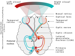
An estimated 40% of the axons in the human brain are devoted to visual function in some way. This complexity and capability is why the visual system is the only sensory system that has a medical specialty (ophthalmology) devoted to it, and nothing else.
Media Attributions
- Unit 6 figure 1 Human Brain © Hugh Guiney adapted by Jim Hutchins is licensed under a CC BY-SA (Attribution ShareAlike) license
- Unit 6 figure 2 Nervous System Overview © Isa.tomanelli adapted by Jim Hutchins is licensed under a CC BY-SA (Attribution ShareAlike) license
- Unit-6-figure-3-canonical-neuron-1 © Jim Hutchins is licensed under a CC BY (Attribution) license
- Unit 6 figure 4 Ear © Lars Chittka; Axel Brockmann - Perception Space—The Final Frontier, A PLoS Biology Vol. 3, No. 4, e137 doi:10.1371/journal.pbio.0030137 (Fig. 1A/Large version), vectorised by Inductiveload is licensed under a CC BY (Attribution) license
- Unit 6 figure 5 Eye Cross Section © Holly Fischer is licensed under a CC BY (Attribution) license
- Unit 6 figure 6 Visual Pathways © Miquel Perello Nieto is licensed under a CC BY-SA (Attribution ShareAlike) license
