The Urinary System
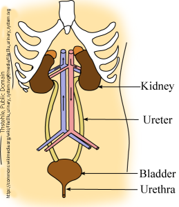
The kidneys filter about 180 liters of our blood per day, producing urine as a waste by-product. Other urinary system structures, including the ureters, bladder, and urethra, provide a drainage and storage system for urine. The urinary system is not simply a waste removal system; it also plays a vital role in maintaining homeostasis by assisting in the control of the body’s acid/base balance (pH), electrolytes, metabolites, fluid volume, and fluid concentration. Each kidney has about a million nephrons, which are the functional units of the kidney that filter, reabsorb, and secrete fluids. A dysfunctional urinary system will have effects on all other systems of the body.
The kidney is a kidney-bean-shaped organ, which probably comes as no surprise to anyone who has seen a kidney or a kidney bean. It has a renal pelvis (the inner curve of the “bean”, root word pyel/o) where the renal artery enters and the renal vein and ureter leave. The slightly darker color in the middle figure shows the renal pyramids, each of which feeds urine into a minor calyx; the urine from the minor calyces drains into a larger major calyx. The major calyces, in turn, join to form the ureter. There is one ureter per kidney, and both the right and left ureter form a functional connection between the kidney and the urinary bladder (root word cyst/o or vesic/o) where urine is stored before it is expelled from the body.
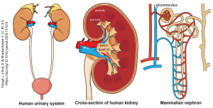
Each nephron consists of a glomerulus (Latin: “little ball of thread”), shaped like a little ball of thread with ample blood supply. That blood is filtered by the glomerulus and the filtrate is collected in the glomerular capsule before it passes into a series of tubes, each of which prepares the filtrate in different ways:
- the proximal convoluted tubule
- the nephron loop (loop of Henle)
- descending limb of nephron loop
- ascending limb of nephron loop
- distal convoluted tubule
- collecting duct
A network of blood vessels (vasa recta) runs parallel to the nephron loop to receive any materials coming out of the nephron loop. Each nephron ends in a single collecting duct, and it is those hundreds of thousands of collecting ducts that come together to form the minor calyx we saw earlier.
The kidney has a major role in the fluid, acid-base, and salt balance of the body. Under the influence of hormones (Unit 7) such as antidiuretic hormone, excess water is secreted by the body. If the blood is too high in solutes (i.e. too salty), then water is retained to reduce its osmolarity. Hydrogen, sodium and potassium ions are actively moved by the kidney either into the nephron loop or into the vasa recta (nearby bloodstream) to keep the blood concentration of these ions constant.
Glucose passes through the glomerular filtration system and into the filtrate within the nephron, and therefore must be replaced to avoid depletion of blood glucose.
Protein metabolism results in the production of urea and other nitrogen-containing (azo) compounds. The production of these wastes is tracked by examining the lab value called blood urea nitrogen (BUN). The kidney is responsible for removing these compounds from the blood and placing them in the urine.
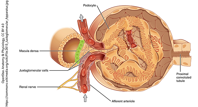
The kidney regulates blood pressure as well. Its own pressure is controlled by receptors in the juxtaglomerular apparatus, which controls the amount of filtration that occurs in the glomerulus. There, an area of the distal convoluted tubule called the “dense spot” (macula densa) joins with juxtaglomerular cells in the lining of the arteriole supplying the nephron with blood to be filtered. This allows detection and regulation of blood pressure, both going into the nephron but also for the body generally. The juxtaglomerular cells secrete the hormone renin, which participates in a complex pathway that ultimately controls blood pressure.
The kidney also plays a key role in blood levels of calcium; it responds to parathyroid hormone (PTH; Unit 7) and vitamin D by increasing retention of calcium to keep blood calcium levels within normal limits.
Media Attributions
- Unit 11 figure 1 Urinary System © Thstehle is licensed under a Public Domain license
- Unit 11 figure 2 Urinary System Kidney cross section nephron © Laura Keogh, David Kilroy, Sourav Bhattacharjee is licensed under a CC BY (Attribution) license
- Unit 11 figure 3 Glomerulus © OpenStax Anatomy and Physiology is licensed under a CC BY (Attribution) license
