The Integumentary (Skin) System
Only [Med Terms] in the Building Episode 4
Corrections: The majority of leather comes from cow skin. The majority of gelatin comes from pig skin.
A comprehensive list of Acme Corporation products is found here.

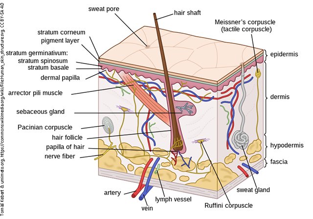
The skin is composed of distinct layers, with the epidermis being the most superficial, the dermis in the middle, and the subcutaneous layer being the deepest of the three. The subcutaneous layer is also called the hypodermis, as shown here. “Hypodermic” is generally used to refer to the needle and “subcutaneous” (abbreviated SC, SQ, or subQ, depending on the clinic) to the layer it is inserted into. Injecting substances with a hypodermic needle is called administering a drug through the subcutaneous (subQ) route. Hair and gland structures are surrounded by a layer of epidermis (red and purple in the diagram) although they appear to be located in the dermis. Blood vessels, lymphatic vessels, and nerves are most abundant in the dermis, but may extend into the subcutaneous layer. The epidermis is made up of many layers of cells and is capable of withstanding the friction our skin encounters on a daily basis.

As the body’s first contact point with the outside world, the skin is exposed to a variety of potentially infectious agents as well as harsh conditions, caustic agents, and other substances that can cause irritation, damage, or disease. The many pathologic conditions of the integumentary system are classified based primarily on appearance and often include reference to the changes in skin color and texture. For example, the normal red appearance seen on the skin as the result of a burn is referred to as erythroderma, simply meaning red skin. The erythroderma shown here is the result of more serious skin disease.
Media Attributions
- Unit 4 figure 1 Skin Diagram © Tomáš Kebert & umimeto.org is licensed under a CC BY-SA (Attribution ShareAlike) license
- Unit 4 figure 2 Erythroderma © DermNet is licensed under a CC BY-NC-ND (Attribution NonCommercial NoDerivatives) license

