Transduction by Metabotropic Receptors
Caleb Bevan
Objective 3: Give examples of signal transduction by metabotropic receptors (G protein-coupled receptors).
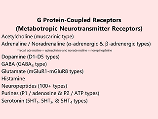
While there are only eight known ionotropic receptors, there are over 130 metabotropic neurotransmitter receptor types. What gives the central nervous system a rich and varied “toolkit” of receptors necessarily creates confusion in trying to understand them all. Here, we will give only two examples. Because they are well-studied, the metabotropic receptors for acetylcholine and GABA will be presented here, so that we can compare and contrast them to the nicotinic acetylcholine receptor and GABAA receptor, ionotropic receptors presented in the previous objective.
The Muscarinic Acetylcholine Receptor
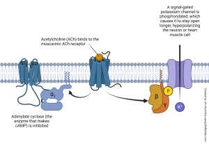
The muscarinic acetylcholine receptor (mAChR), like most G protein-coupled receptors, is a protein with seven transmembrane helical regions. When activated, the associated G protein splits apart. The α subunit of the G protein inhibits adenylate cyclase (adenylyl cyclase) and reduces the amount of cAMP produced in the cell.
The β and γ subunits of the G protein, acting together, add phosphate groups onto a signal-gated potassium channel. When phosphorylated, the K+ channel is more likely to be open, which means that K+ leaves the cell, making the cell more negative (hyperpolarization) and driving the membrane voltage closer to the equilibrium potential for potassium (EK). The overall effect is to inhibit the target cell. For example, mAChRs on heart muscle slow the heartbeat by driving the heart muscle cell membrane potential downwards, and it takes longer for the heart muscle to reach the action potential threshold. This longer climb to threshold slows the heartbeat.
The GABAB Receptor
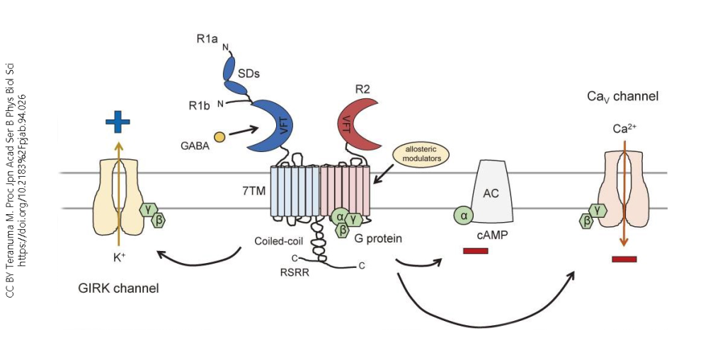
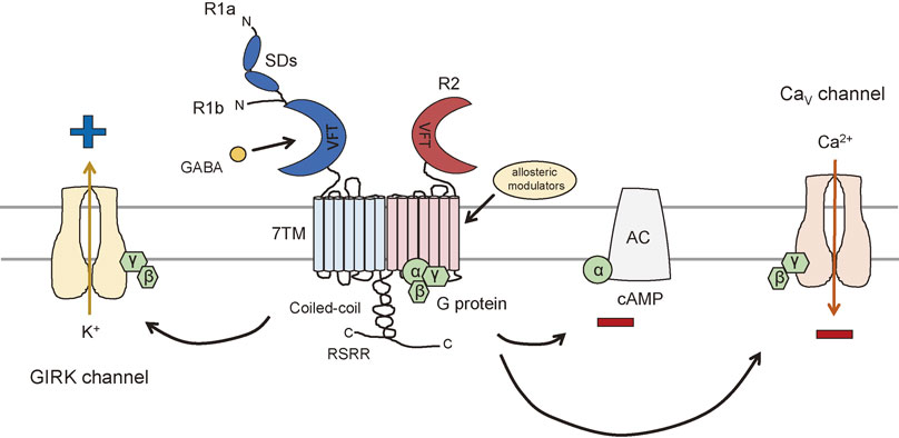
Like the mAChR and the GABAA receptor, the GABAB receptor, when activated, causes inhibition of neuronal responses. The receptor has seven transmembrane (7TM) regions, like most GPCRs. When the receptor is bound by GABA or another ligand, the associated G protein splits, and the α subunit inhibits adenylate cyclase. The β and γ subunits phosphorylate the G protein-coupled inwardly rectifying potassium (GIRK) channel, causing it to be more likely to open. The potassium outflow hyperpolarizes the neuron. (Inward rectification is an electrical property of some channels and will be discussed in a more advanced cell and molecular neuroscience class, if you decide to take one.)
The β and γ subunits also act on the voltage-gated Ca2+ channel, making it less likely to open and admit Ca2+ into the neuron.
Both activation of the GIRK channel and inhibition of the voltage-gated Ca2+ channel make neurotransmitter release less likely, and so like the mAChR and the GABAA receptor, the GABAB receptor is classified as an inhibitory receptor.
Media Attributions
- GCPR table © Jim Hutchins is licensed under a CC BY-SA (Attribution ShareAlike) license
- Muscarinic acetylcholine receptor © Jim Hutchins is licensed under a CC BY-NC-ND (Attribution NonCommercial NoDerivatives) license
- GABA-B receptor © Miho Terunuma is licensed under a All Rights Reserved license
