Spatial and Temporal Summation
Objective 8: State the principles underlying spatial and temporal summation of graded potentials.
Each of the 10,000 inputs onto a neuron is weighted differently.
Each neuron has at least one decision point where the neuron asks itself: Do I fire an action potential, or not? Do I release neurotransmitter, or not?
From the perspective of the decision point, the sum total of the weighted inputs either results in a voltage above threshold, or it doesn’t. That is how the question is answered. At this location, is my membrane voltage above threshold, or not? If it is, then an action potential is generated at the axon hillock. If it is, then neurotransmitter is released from the active zone of a synapse.
Each EPSP and IPSP in a neuron is termed a graded potential. (Remember that “potential” and “voltage” are the same thing.) As we saw in Objective 6, graded potentials decrement in space following mathematical rules that are invariant. The larger the distance between the source of the voltage change and the decision point (i.e. the more length constants the graded potential has to traverse), the smaller the voltage change becomes. The more time that passes after the voltage change, the smaller the voltage change becomes.
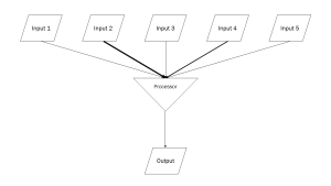 In this diagram, which is deliberately designed as a computer flowchart, with standardized symbols: a parallelogram for inputs (postsynaptic potentials) and a triangle for a merge point. This represents where the dendritic spines receive EPSPs or IPSPs, which then pass into small dendritic branches that come together at a single point. Note the weight of the arrow is different, because PSPs come in different sizes, and different dendritic branches have different calibers and therefore different length and time constants. The EPSPs and IPSPs are all summed together depending on their size and direction at the branch point (triangle in this diagram). This becomes the output of this system of branches.
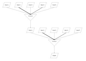
 Imagine that five of these tiny branches gather together into the next branch point. Only one is shown, but you can see that the output (summed voltage) of each branch point (1, 2, 3, 4, 5) becomes the input voltage of the new branch point. This continues, with each dendritic tree, until finally the ultimate output (potential, voltage) reaches a decision point and threshold is reached — or not.
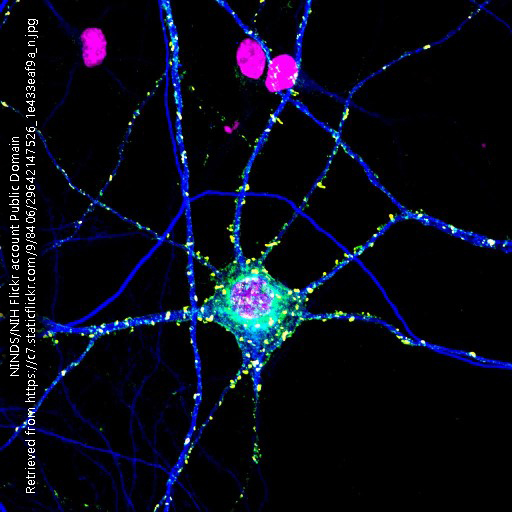
To help you visualize this branching, imagine EPSPs and IPSPs coming in at each of the dendritic spines (bumpy shapes) on this retinal bipolar cell. Then imagine the voltages summing together at each branch point, and that summed voltage moving forward to the next branch point, and so on.
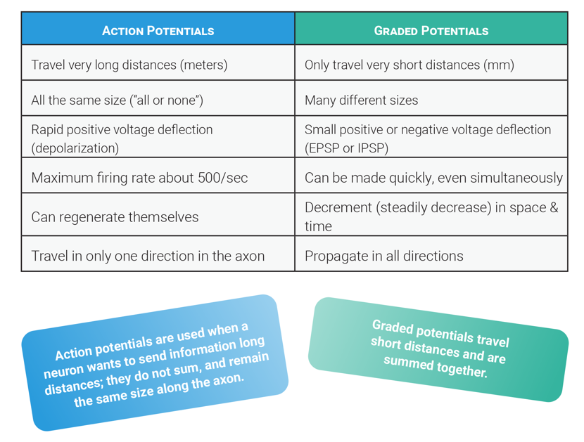
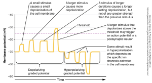 Because graded potentials come in different sizes, as shown here, they can be summed together over distance and in time. Imagine what happens to each of the PSPs shown as they travel over distance, or as time passes.
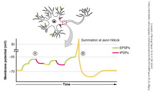 The closer a postsynaptic potential is to the decision point, the greater the influence it has on the eventual decision being made. This concept is called spatial summation. A superthreshold postsynaptic potential which is right next to the active zone of a synapse almost certainly will cause neurotransmitter to be released. An input to a projection neuron at the axon hillock, if large enough to open enough voltage-gated Na+ channels, will almost certainly cause an action potential to be generated.
The more closely postsynaptic potentials are spaced in time, the larger the eventual sum when they are added together. This concept is called temporal summation. Many small EPSPs all arriving at a decision point at the same time will be likely to result in a superthreshold stimulus. If, as shown in the diagram, there is 15 mV between the resting potential and the threshold, then sixteen 1 mV stimuli all arriving at the same time will result in an action potential. If, however, there is a 1 msec space between each, then each one will decrement in time and the chance of triggering an action potential is now quite small.