Postsynaptic Potentials
Caleb Bevan
Objective 7: Describe how the events on the postsynaptic side of the synapse result in postsynaptic potentials.

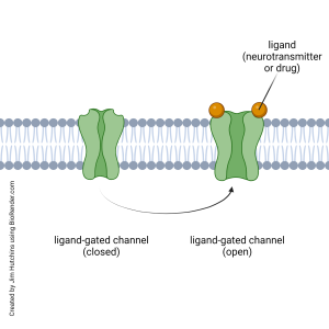
When neurotransmitters bind to ionotropic receptors, as shown here at left, a ligand-gated channel opens or closes.
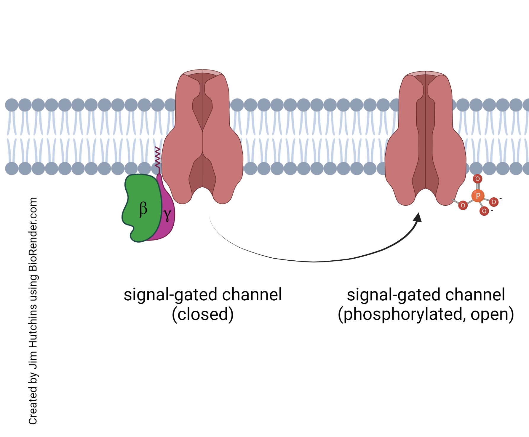
 When neurotransmitters bind to a G protein-coupled receptor, as shown here at right, one possible effect is to create an intracellular signal that opens or closes a signal-gated ion channel.
Either way, there is a change in the flow of ions across the neuronal membrane. There are only four ions which move across the cell membrane in any quantity: Na+, K+, and Ca2+ (cations, positively-charged ions) and Cl– (an anion, a negatively-charged ion). When we divide these into cations and anions, and consider flow either into a neuron or out of it, only four possibilities pertain:
- When cations flow into a neuron through an ion channel, the cell is depolarized.
- When cations flow out of a neuron through an ion channel, the cell is hyperpolarized.
- When anions flow into a neuron through a chloride channel, the cell is hyperpolarized.
- When anions flow out of a neuron through a chloride channel, the cell is depolarized.
The small change in voltage (depolarization or hyperpolarization) that occurs as a result of synaptic action is called the postsynaptic potential.
If the postsynaptic potential moves the membrane voltage closer to threshold, and thereby makes an action potential or synaptic release more likely to happen, it is called an excitatory postsynaptic potential (EPSP).
If the postsynaptic potential moves the membrane voltage further from threshold, and thereby makes an action potential or synaptic release less likely to happen, it is called an inhibitory postsynaptic potential (IPSP).
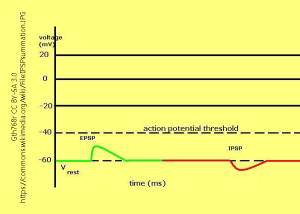
 Examples of EPSPs and IPSPs are shown on the graph. Note the position of the action potential threshold, which is the value at which there are enough voltage-gated Na+ channels opening to make the rise in membrane potential self-sustaining (i.e., an action potential). A similar argument can be made for the threshold to trigger enough voltage-gated Ca2+ channels to cause synaptic release.
The EPSP moves the membrane potential closer to threshold; the IPSP moves the membrane potential further away from threshold. It is rare for a single EPSP to be large enough to trigger an action potential or synaptic release.
Media Attributions
- Generic ligand gated channel function © Jim Hutchins is licensed under a CC BY-NC-ND (Attribution NonCommercial NoDerivatives) license
- Generic signal-gated channel function © Jim Hutchins is licensed under a CC BY-NC-ND (Attribution NonCommercial NoDerivatives) license
- Postsynaptic potentials © User:Gth768r adapted by Jim Hutchins is licensed under a CC BY-SA (Attribution ShareAlike) license

