Optic Nerve and Tract
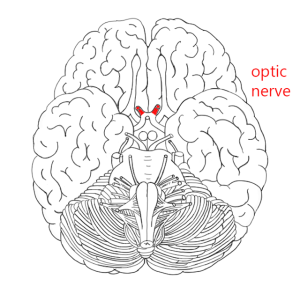 The optic nerve carries all the axons from the retinal ganglion cells. Recall that we saw this structure as the optic nerve head in the retina producing the blind spot in visual perception.
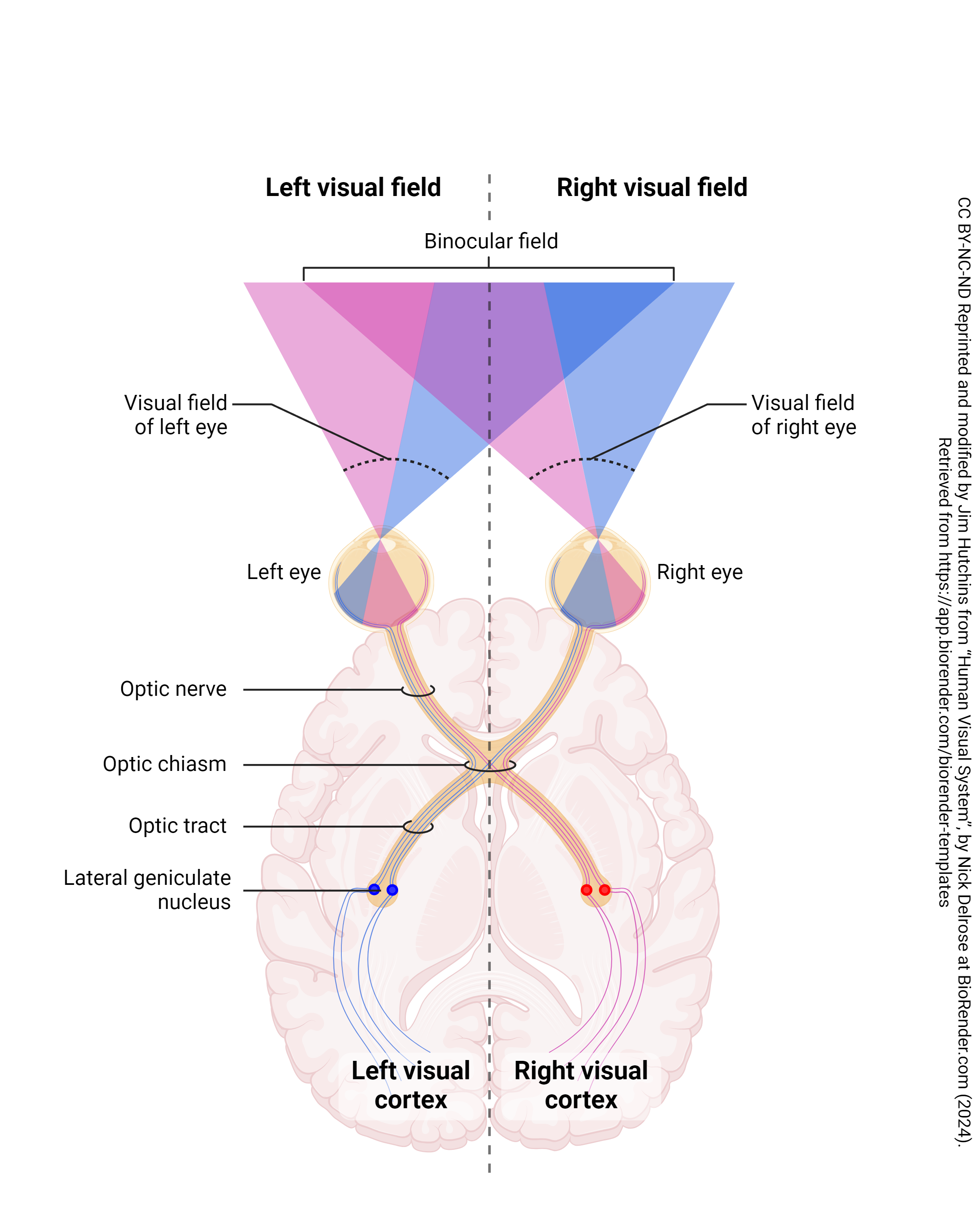
Half of the axons from each eye, representing the right temporal retina and the left nasal retina, are picking up information from the left visual world. These are represented by pink shading and pink axons.
Half of the axons from each eye, representing the right nasal retina and the left temporal retina, are picking up information from the right visual world. These are represented by blue shading and blue axons.
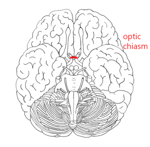
Thus, at the optic chiasm (the Greek letter chi, χ, looks like this structure), the axons from the nasal half of each retina cross to join those that carry information from the same part of the visual field.
When something is on the same side in neuroscience, we call it ipsilateral. When it’s on the opposite side, we call it contralateral.
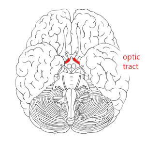 The optic tract, then, contains two complete representations of the contralateral visual field, one from ganglion cell axons originating in the right eye and one from ganglion cell axons originating in the left eye.
For example, the right optic tract contains axons from the left (contralateral) nasal retina ganglion cells and the right (ipsilateral) temporal retina ganglion cells.
