Myelination
Jim Hutchins
Objective 4: State the purpose of myelination and its main features.
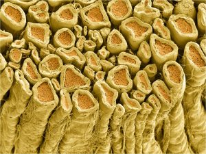
Some axons have an insulating wrapping surrounding them. We will see the function of this wrapping later, but for now, all you need to know is that myelin improves the speed and fidelity of information flow down the axon, from the cell body to the synaptic terminal. There is a difference between the myelin wrapping in the CNS, which does not support axonal regrowth in case of injury, and myelin wrapping in the PNS, which does. Both kinds of myelin are mostly (80%) lipid, but PNS myelin has specialized protein (like recognition and adhesion proteins) which supports axonal regrowth.
Accordingly, it’s a different cell type that makes myelin in the CNS (oligodendrocytes) and PNS (Schwann cells).
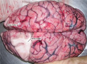
 Myelin, like most lipids, appears white when you cut into it. This is the basis for so-called white matter and gray matter in the brain. (It’s really less-pink-matter and darker-pink-matter in a freshly cut or living brain, but those aren’t the terms we are given.)
White matter is rich in myelinated axons and generally indicates the presence of one or more fiber tracts. Remember a tract is a bundle of neuronal axons traveling to and from the same place.
Gray matter is rich in cell bodies and dendrites. Because those are (in general) the processing parts of the neuron, most processing is done in the gray matter.

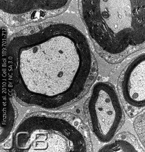
Because the phospholipid head groups and proteins found in myelin avidly bind to osmium tetroxide, in the electron microscope, myelinated axons appear to have a dense, jet-black wrapping surrounding them. This dense layer of insulating lipid will not allow many ions to cross.

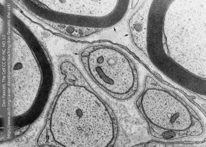
Myelinated axons carry this thick myelin sheath. Unmyelinated axons are not without protection, however. Here, we see a single Schwann cell process enrobing three unmyelinated axons in a peripheral nerve.

The myelin wrapping around nerves starts out as a thin extension of the oligodendrocyte or Schwann cell. In keeping with the name oligodendrocyte (“several branches cell”), there are several such thin processes that each touch a different axon. Once it touches the axon, it starts to crawl in a circular pattern over and over and over again, until several dozen layers of membrane and a thin sheet of cytoplasm surround each axon. The cytoplasm and any remaining extracellular fluid are slowly squeezed out, like toothpaste from a tube, until all that remains are lipids with the proteins which screen off negative charges and allow for cellular recognition.
Media Attributions
- Myelinated axons © Deerinck, Tom and Ellisman, Mark/ National Center for Microscopy and Imaging Research (NCMIR) is licensed under a CC BY-NC (Attribution NonCommercial) license
- White and Gray Matter © Betts, J. Gordon; Young, Kelly A.; Wise, James A.; Johnson, Eddie; Poe, Brandon; Kruse, Dean H. Korol, Oksana; Johnson, Jody E.; Womble, Mark & DeSaix, Peter is licensed under a CC BY (Attribution) license
- Myelinated and unmyelinated axons © Markus Finzsch, Silke Schreiner, Tatjana Kichko, Peter Reeh, Ernst R. Tamm, Michael R. Bösl, Dies Meijer, Michael Wegner adapted by Jim Hutchins is licensed under a CC BY-NC-SA (Attribution NonCommercial ShareAlike) license
- Cell Surface © Don W. Fawcett is licensed under a CC BY-NC-ND (Attribution NonCommercial NoDerivatives) license
- Myelination © Betts, J. Gordon; Young, Kelly A.; Wise, James A.; Johnson, Eddie; Poe, Brandon; Kruse, Dean H. Korol, Oksana; Johnson, Jody E.; Womble, Mark & DeSaix, Peter adapted by Jim Hutchins is licensed under a CC BY (Attribution) license

