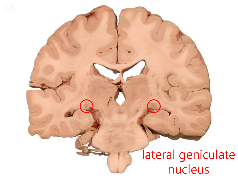
Lateral Geniculate Nucleus (LGN)


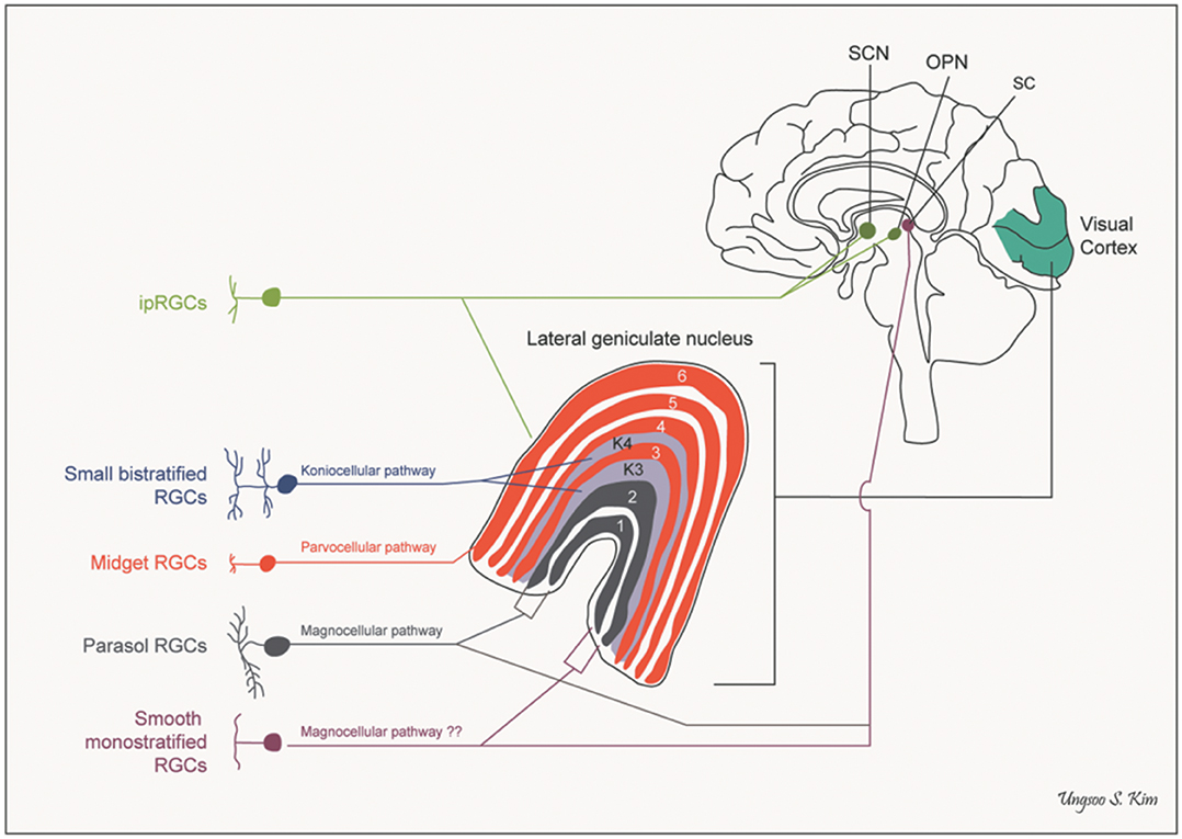
There are six layers of cells in the human LGN. Two (termed, naturally, layers 1 and 2) contain large cells and are called the magnocellular layers. Four (layers 3, 4, 5, and 6) contain smaller cells and are called the parvocellular layers. Late in our study of the LGN, we found that much of the action was occurring in even smaller cells in the LGN scattered throughout and between the layers but we had run out of names for sizes (magno– = “large”; parvo– = “small”) so in desperation, we called those koniocellular cells from the Greek konio– = “dust”.
Layers 1, 4, and 6 are innervated by ganglion cell axons from the contralateral side. Layers 2, 3, and 5 are innervated by ganglion cell axons from the ipsilateral side. M ganglion cells terminate in the magnocellular layers and P ganglion cells terminate in the parvocellular layers. (Non-M, non-P ganglion cells terminate onto the koniocellular cells.) So, in summary,
- Magnocellular layers
- 1: contralateral M parasol ganglion cells
- 2: ipsilateral M parasol ganglion cells
- Parvocellular layers
- 3: ipsilateral P midget ganglion cells
- 4: contralateral P midget ganglion cells
- 5: ipsilateral P midget ganglion cells
- 6: contralateral P midget ganglion cells
Each layer, then, contains a complete representation of objects in the contralateral visual field, with the same point in visual space “stacked” on top of a cell that conveys information about the same point. Like a toothpick in a layer cake, the same point in visual space is represented six times in the six layers of the LGN.

