Autonomic Control of Heart Rate
Jim Hutchins
Objective 4: Recognize the mechanisms by which heart rate is controlled in the autonomic nervous system.
Let’s turn to a specific and important example: the innervation of the heart.
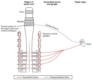
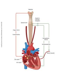
Recall that cranial nerve X, the vagus nerve, is named this because it wanders all over the thoracic and abdominal cavities. Therefore, cell bodies of the parasympathetic nervous system which innervate the heart are located in the dorsal motor nucleus of the vagus and the nucleus ambiguus. Their axons travel through the vagus nerve to reach the heart. Parasympathetic (“rest and digest”) action is to slow the heart, and it does this by releasing acetylcholine onto the pacemaker cells in the sinoatrial node of the heart.
For the sympathetic innervation of the heart, preganglionic cell bodies are found in the lateral horn of the spinal cord (green region of the diagram) at upper thoracic levels (there is some variability, but T1-T5 would be a good estimate). These send out axons which synapse in the T1-T5 sympathetic chain ganglia but also in some chain ganglia not directly linked to the spinal cord: the superior cervical ganglion, middle cervical ganglion, and inferior cervical ganglion. All these ganglia contain postganglionic cell bodies of neurons whose axons travel in the cardiac accelerator nerve to the pacemakers of the heart, where they release norepinephrine (noradrenaline) to speed up the heart. This is exactly what you want to do in a sympathetic (“fight or flight”) response.
Media Attributions
- Heart autonomics © Betts, J. Gordon; Young, Kelly A.; Wise, James A.; Johnson, Eddie; Poe, Brandon; Kruse, Dean H. Korol, Oksana; Johnson, Jody E.; Womble, Mark & DeSaix, Peter is licensed under a CC BY (Attribution) license
- Sympathetic Reflexes in the Heart © Emma Cornelius is licensed under a CC BY-NC-ND (Attribution NonCommercial NoDerivatives) license
