The Pupillary Light Reflex
Jim Hutchins
Objective 5: Describe the circuitry which controls pupil size.
Brainstem Nuclei Controlling Pupil Size

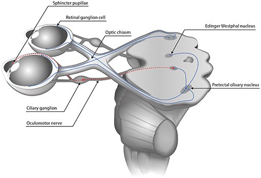
Because the eye must control the amount of light reaching the retina, so that it is roughly equal in bright sunlight or dim interior rooms, the midbrain nuclei controlling pupil size must receive some input from the retinal ganglion cells which transmit information about overall light levels. Recall that these melanopsin intrinsically photosensitive ganglion cells have receptive fields that cover the entire retina. Axons of these large-receptive-field ganglion cells project first to the pretectal olivary nucleus (PON) of the midbrain, and the OPN in turn sends short axons to the nearby Edinger-Westphal nucleus, the autonomic motor nucleus which controls pupil size. The olivary pretectal nucleus is called the olivary pretectal nucleus by some authors.
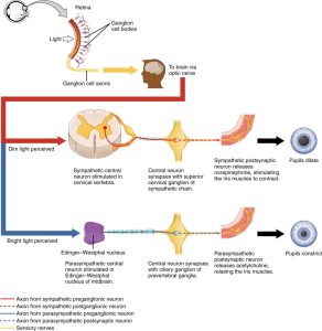
The pupil is controlled by the sympathetic and parasympathetic nervous system.
When the sympathetic response dilates or enlarges the pupil (mydriasis), norepinephrine is released onto the pupillary dilator muscle. This smooth muscle runs radially from the center of the pupil, so contraction of its fibers increases the diameter of the pupil.
When the parasympathetic response constricts or shrinks the pupil (miosis), acetylcholine is released onto muscarinic ACh receptors of the pupillary sphincter muscle. These muscle cells run in a band around the edge of the pupil, so contraction of its fibers decreases the diameter of the pupil. Belladonna or atropine, remember, are drugs that block these muscarinic ACh receptors and allow the dilator muscle to act unopposed.
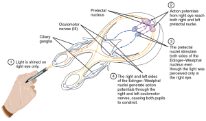
When we shine a light into the eye, the signal is carried by the optic nerve (CN II). Most of these ganglion cell axons end in the lateral geniculate nucleus of the thalamus but about 10% continue to the midbrain where they mediate visual reflexes like looking at a flash of light or the pupillary light reflex we’re discussing now. In this case, the axons end in the Edinger-Westphal nucleus, which is the name for the cluster of parasympathetic motor neuron cell bodies which lie deep in the midbrain. These preganglionic axons extend out to the ciliary ganglion where they synapse onto postganglionic neurons that travel a short distance into the iris itself. There, their axon terminals release acetylcholine onto muscarinic receptors, causing the pupil to shrink (miosis).
In this way we can quickly test the function of the midbrain. Extensive brain damage can affect the midbrain, so the absence of a normal pupillary light reflex is a very grave clinical sign.
Innervation of the Iris: Controlling Pupil Size
We’ll take a detailed look at another organ, the iris (colored part of the eye) which defines a hole in the middle called the pupil. As you’ve probably discovered, the pupil gets larger and smaller in response to light (reaction) but also in response to focusing (accommodation) and emotional events.
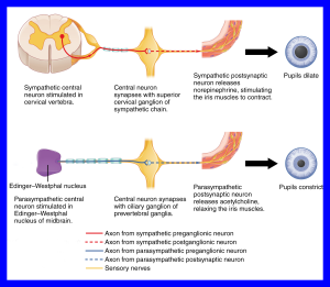
The sympathetic nervous system causes the pupil to dilate. You can easily confirm this by pissing off your cat or your partner. (Probably best not to piss off both at the same time.) The circuitry for this response is as follows:
- Preganglionic neurons are found in the lateral horn of the upper thoracic spinal cord
- Their axons release acetylcholine onto cells in the superior cervical ganglion
- The postganglionic axons take an ill-defined, meandering course along blood vessels to the oculomotor nerve (CN III)
- The axon terminals release norepinephrine (noradrenaline) onto the iris dilator muscle, smooth muscle fibers that run radially in the iris to cause it to enlarge the pupil (mydriasis)
For the parasympathetic innervation of the iris:
- Preganglionic neurons are found in the midbrain
- Their axons release acetylcholine onto cells in the ciliary ganglion, near the eye
- The postganglionic axons travel in the oculomotor nerve (CN III) a short distance to reach the iris
- There, they release acetylcholine onto the iris sphincter muscle, smooth muscle fibers which run circumferentially around the rim of the pupil and cause it to shrink (miosis)
 In medieval Italy, women got the idea that men were so stupid that they could be made to fall in love with you just because your pupils were dilated — and therefore men responded to simple autonomic responses indicating the woman was actually, you know, interested in what he had to say. To achieve this state, they would take a drug called belladonna, extracted from the deadly nightshade Atropa belladonna. (“Belladonna”, in Italian means literally, “beautiful woman” even if she’s not named “Donna”.)
You probably figured out already how dan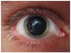 gerous this is, because we call the plant the deadly nightshade after all. It’s much safer to use a purified form of belladonna called atropine. Atropine is in everyday use in the clinic. For example, you’ve probably been given atropine eye drops to dilate your pupils during an eye exam. Atropine is also used to regulate the heartbeat. Now we understand how this works: atropine blocks the receptors for acetylcholine in the heart, causing it to speed up. Atropine also blocks the receptors that make the pupil small in the iris sphincter muscle, causing it to become bigger.
Media Attributions
- Pupillary pathway © Carina Kelbsch, Torsten Strasser, Yanjun Chen, Beatrix Feigl, Paul D. Gamlin, Randy Kardon, Tobias Peters, Kathryn A. Roecklein, Stuart R. Steinhauer, Elemer Szabadi, Andrew J. Zele, Helmut Wilhelm, and Barbara J. Wilhelm is licensed under a CC BY (Attribution) license
- Reflex pupil © Betts, J. Gordon; Young, Kelly A.; Wise, James A.; Johnson, Eddie; Poe, Brandon; Kruse, Dean H. Korol, Oksana; Johnson, Jody E.; Womble, Mark & DeSaix, Peter is licensed under a CC BY (Attribution) license
- Pupil reflex pathway © Betts, J. Gordon; Young, Kelly A.; Wise, James A.; Johnson, Eddie; Poe, Brandon; Kruse, Dean H. Korol, Oksana; Johnson, Jody E.; Womble, Mark & DeSaix, Peter is licensed under a CC BY (Attribution) license
- Pupil autonomics © Betts, J. Gordon; Young, Kelly A.; Wise, James A.; Johnson, Eddie; Poe, Brandon; Kruse, Dean H. Korol, Oksana; Johnson, Jody E.; Womble, Mark & DeSaix, Peter adapted by Jim Hutchins is licensed under a CC BY (Attribution) license
- Belladonna © Plbmak is licensed under a CC BY-NC-ND (Attribution NonCommercial NoDerivatives) license
- Mydriasis © Betts, J. Gordon; Young, Kelly A.; Wise, James A.; Johnson, Eddie; Poe, Brandon; Kruse, Dean H. Korol, Oksana; Johnson, Jody E.; Womble, Mark & DeSaix, Peter adapted by Jim Hutchins is licensed under a CC BY (Attribution) license
