Sensory System: The Visual System
The visual system is probably the most-used and most important of the human sensory systems. Some authorities estimate that about 40% of the human CNS is devoted to receiving and processing visual information.
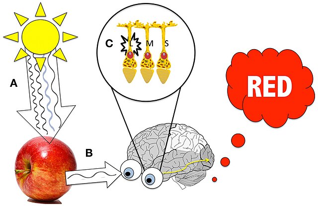
The eye is the sensory end-organ responsible for receiving visual information. The eye is responsible for refracting light rays so that an image is focused on the retina. The retina is an outpocketing of the central nervous system with neurons that are responsible for transducing photons into an electrochemical signal that may be used by the central nervous system. The cells responsible for transduction are called photoreceptors.
The retina also contains neurons that process the visual signal and prepare the signal for processing by the remainder of the visual system. These cells are called horizontal cells, bipolar cells, and amacrine cells. The synaptic processing converges on the dendrites of ganglion cells.
Ganglion cells are the output cells of the retina. Their axons form the optic nerve, which rather than a true nerve (which is the name of a bundle of axons that make up part of the peripheral nervous system) is a bundle of ganglion cell axons making up part of the central nervous system. The optic nerves converge at the optic chiasm (named because of its shape like the X-shaped Greek letter chi); some axons remain on the same side and some cross, and the resorted axons continue as the optic tract.
Ganglion cell axons terminate in the lateral geniculate nucleus (LGN, also called the LGB or lateral geniculate body), which is part of the thalamus. After further processing of the visual signal in the LGN, the axons of LGN relay cells form the optic radiations whose axons terminate in layer IVc in the cortex of the occipital lobe.
Unfortunately, the area where visual information is processed next is called by four different names which are used interchangeably by various authorities. Since we can’t say which sources you’ll be reading, we have to give you all four names.
- Primary visual cortex (1° visual cortex) is the name given to the occipital lobe regions where the axons of LGN relay cells terminate, and where the visual signal undergoes a series of elaborate processing steps. This naming convention pays homage to the standard way of naming sensory cortex; the place in the brain where sensory information first reaches consciousness is called the primary [name of sensory system] cortex. For example, there is a primary auditory cortex and primary somatosensory cortex.
- Striate cortex is a name given to the primary visual cortex because the myelinated axons of millions of LGN neurons form a macroscopically visible stripe (stria) in the occipital cortex. The 18th century Italian anatomist Francesco Gennari was the first to describe this area.
- Brodmann area 17 is the designator given by Korbinian Brodmann in his 1909 map of cytoarchitectonic areas of cortex.
- The calcarine sulcus is the deep groove which runs through the center of visual cortex. The word “calcar” means a raptor’s claw, which it was thought to resemble.
These names are used interchangeably in this book, just as they are in the literature.
A large swath of the parietal and temporal lobes are also devoted to increasingly complex levels of visual analysis, as we will discuss later.
Objectives for the Visual System include:
- Explain how the gross anatomy of the eye contributes to the initial processing of the visual signal.
- Discuss the microscopic anatomy of the retina.
- Relate how the three cone types of the human retina contribute to color vision.
- State the processes involved in transducing a photon into a change in neurotransmitter release in photoreceptors.
- Describe how each of the five neuronal types of the retina contribute to visual processing.
- Explain how retinal bipolar cells act as edge detectors.
- Summarize how amacrine cell and bipolar cell inputs to ganglion cells mediate motion processing.
- Demonstrate how retinal ganglion cells process information to produce a neural output signal from the retina.
- Diagram the visual fields.
- Describe the retinofugal pathway to the lateral geniculate nucleus, midbrain, and hypothalamic nuclei.
- Recognize key features of the pathway between the lateral geniculate nucleus and visual cortex (the optic radiations).
- Recognize the location and basic cytoarchitectonic organization of visual cortex.
- Construct a model for binocular vision (stereopsis).
- Discuss the organization of visual cortex into cortical modules (hypercolumns).
- Describe the anatomy and function of the dorsal and ventral pathways for visual information.
Media Attributions
- Seeing red © A Wade & A Benjamin is licensed under a CC BY (Attribution) license
