Urination
Jim Hutchins
Objective 8: Describe the pathways that control urination (micturition).
Urination is the “U” in the mnemonic SLUDD which is used to remember the function of the parasympathetic nervous system. Urination is also called micturition, which is a word that describes the active process of urination.

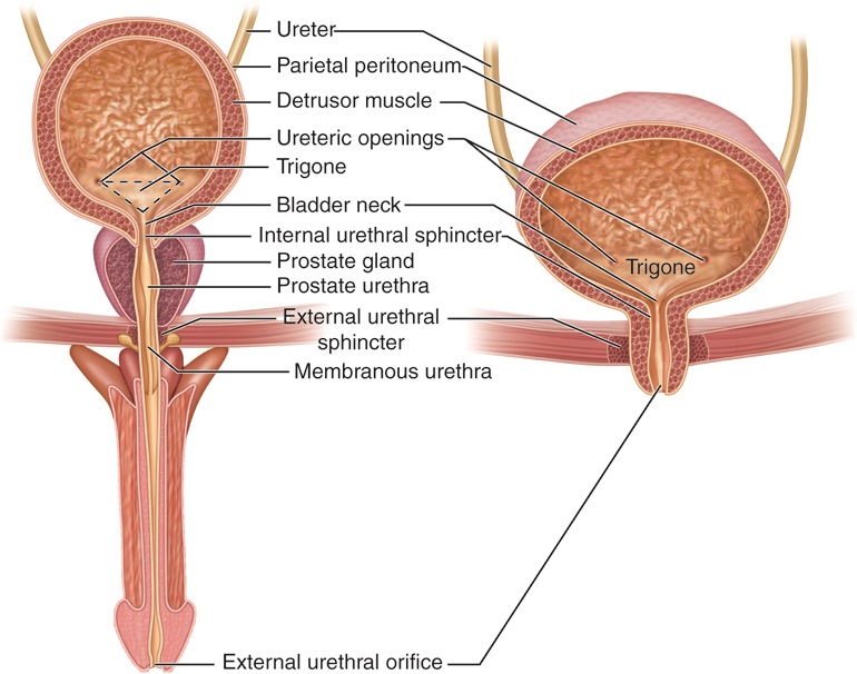
The detrusor muscle is the name for overlapping bands of smooth muscle that surround the urinary bladder. When the detrusor muscle is relaxed, the bladder can fill with urine. When the detrusor is contracted, the bladder decreases in size and urine is forced out (if allowed to by the sphincter muscles).
The internal urinary sphincter (internal urethral sphincter) is smooth muscle under autonomic control.
The external urinary sphincter (external urethral sphincter) is skeletal muscle, part of the pelvic floor muscle group. It is under voluntary, conscious control.
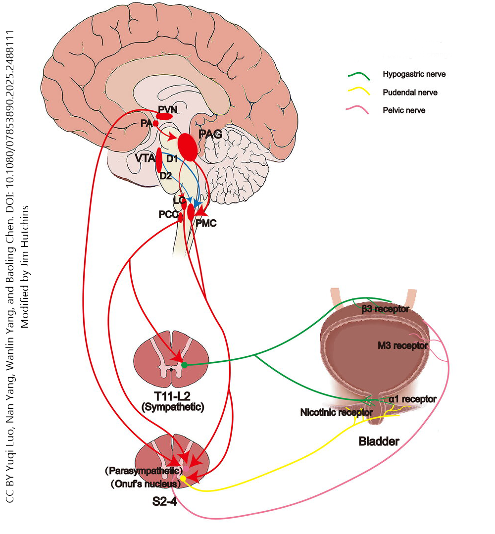
Sympathetic Innervation of the Bladder
The sympathetic nervous system, in general, relaxes the detrusor muscle and inhibits urination.
The short preganglionic cell bodies are found in the lateral (intermediate) horn of the spinal cord at levels T11, T12, L1, and L2. These synapse on postganglionic neuron cell bodies found in the most inferior set of sympathetic chain ganglia. These long postganglionic axons run in the hypogastric nerve, ending in presynaptic contacts in the bladder wall. These neurons release noradrenaline (norepinephrine) onto the detrusor muscle and internal urinary sphincter muscle.
The β3 adrenergic receptors increase the activity of adenylate cyclase, which in turn increases the second messenger cAMP. This relaxes the detrusor muscle.
At the same time, α1 adrenergic receptors of the internal urinary sphincter cause contraction of this muscle and retention of urine in the bladder.
Voluntary Motor Innervation of the Bladder
Voluntary control of micturition is exerted through neurons in the ventral horn of the spinal cord at levels S2, S3, and S4. These form a histologically distinct population of α motor neurons (lower motor neurons) called Onuf’s nucleus. The axons arising from the cell bodies in Onuf’s nucleus form the pudendal nerve and innervate the external urinary sphincter muscle, which is part of the pelvic floor muscle group.
The pons, periaqueductal gray, and thalamus are wired together to exert unconscious control over urination, while the insula, prefrontal cortex, anterior cingulate cortex, and supplementary motor area all contribute to the conscious control of urination. These areas all send axons to Onuf’s nucleus to control urination and ensure that it occurs at socially acceptable times and places.
Parasympathetic Innervation of the Bladder
Preganglionic neuronal cell bodies of the parasympathetic nervous system controlling urination are found in the lateral (intermediate) horn of the spinal cord at levels S2, S3, and S4. These cause smooth muscle contraction of the detrusor muscle via M3 receptors on cells that make up the intramural ganglia found in the wall of the detrusor muscle. These are also activated by stretch receptors in the bladder wall.
Media Attributions
- Urinary structures © Cenveo adapted by Jim Hutchins is licensed under a CC BY (Attribution) license
- Control of urination © Yuqi Luoa, Nan Yangb, Wanlin Yanga, Baoling Chena, Shuzhen Zhua, YihRu Wuc and Qing Wanga adapted by Jim Hutchins is licensed under a CC BY (Attribution) license
