The Hippocampal Slice
Caleb Bevan
Objective 2: Explain the importance of the hippocampal slice preparation to studies of learning and memory.
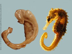
The name hippocampus derives from the Greek word for “seahorse”. It is easy to see how the 16th century Italian anatomist Giulio Cesare Aranzio coined the term: this part of the brain looks like a seahorse.
 It is an important part of the limbic lobe, a set of interconnected structures that make up the bulk of the brain structures both creating and regulating emotional responses. Its inputs come from olfactory and emotional parts of the brain. The cingulate cortex, temporal lobe cortex, amygdala, orbital cortex, and olfactory bulb all feed into the entorhinal cortex, adjacent to the hippocampus. Sensory association areas also connect to the entorhinal cortex. Sensory association areas are the regions of the brain where sensory information is integrated between senses and subjected to higher-order processing which forms a complete, conscious perception of the world. The medial septum, which has been called “an information hub for social memory”, also provides significant input to the hippocampus. A region called the subiculum lies between the entorhinal cortex and the hippocampus.
 The hippocampus, in turn, sends efferents to the entorhinal cortex and amygdala, and via a major fiber bundle called the fornix to the mammillary bodies of the thalamus as well as many areas of cortex. This wiring supports its primordial function: to add an emotional “tag” onto memories of a particular place. One can easily imagine that an animal needs to remember where one encounters dangerous stimuli; I have a clear memory of a specific location on a running trail where I encountered a skunk almost 10 years ago.
The hippocampus is made up of two interlocking letter Cs: one C is called the dentate gyrus. The dentate gyrus, which can be seen in the photograph of the dissected hippocampus, looks like a set of teeth from the outside of the hippocampus, hence the name.
The other letter C has four regions. The entire area of the C has been named cornu Ammon, or “Ammon’s horn” in English, after an Egyptian/Greek deity, named Amun or Amun-Ra in Egypt who became Zeus in Greek mythology. Amun/Ammon was often represented as having a man’s head with ram horns, and those horns are the cornu Ammon. In the hippocampus, cornu Ammon (CA) is divided into four numbered areas: CA1, CA2, CA3, and CA4. CA1 is closest to the subiculum while CA4 is closest to the dentate gyrus.
Of course, Scoville’s inadvertent experiment on Henry Molaison also provided us with important clues as to the function of the human hippocampus.
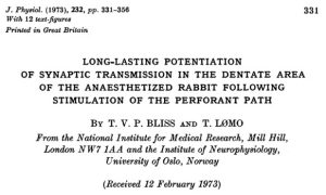 Some 20 years after Henry’s surgery, and 24 years after Hebb’s seminal monograph on modifiable synapses, Timothy Bliss and Terje Lømo published a key paper in the Journal of Neurophysiology describing a new way of studying the hippocampus in experimental animals.
Oxygen, needed for neuronal function, can only penetrate about 1/5 of a millimeter (200 μm) into the tissue from the nearest surface. Normally, the capillaries of the brain which supply oxygen are within this distance of any neuron in the brain. When the hippocampus or any other brain part is removed from its blood supply, however, the researcher must supply oxygen and nutrients in a buffered salt water bath. (Buffered salt water as a blood replacer in physiological experiments is called Ringer’s solution.)
If the dissected tissue is thicker than about 400 μm (i.e., if the center of the slice is more than 200 μm from the surface), it cannot survive for long. By happy accident, the hippocampus is shaped like a jelly roll and all the circuitry one needs to study the hippocampus is located within a single plane of section. Bliss and Lømo sliced the “jelly roll” into sections about 400 μm thick, and were able to use electrophysiological recordings to study the circuitry of the slice as they kept it alive for 24 hours or more.
Bliss and Lømo described a phenomenon in their hippocampal slices which we now call long-term potentiation (LTP). This was immediately recognized as the first electrophysiological demonstration of a Hebbian synapse. After a train of action potentials was triggered in one of the incoming circuits of the hippocampus, the activity of the postsynaptic cell was persistently elevated for as long as they could keep the slice alive.
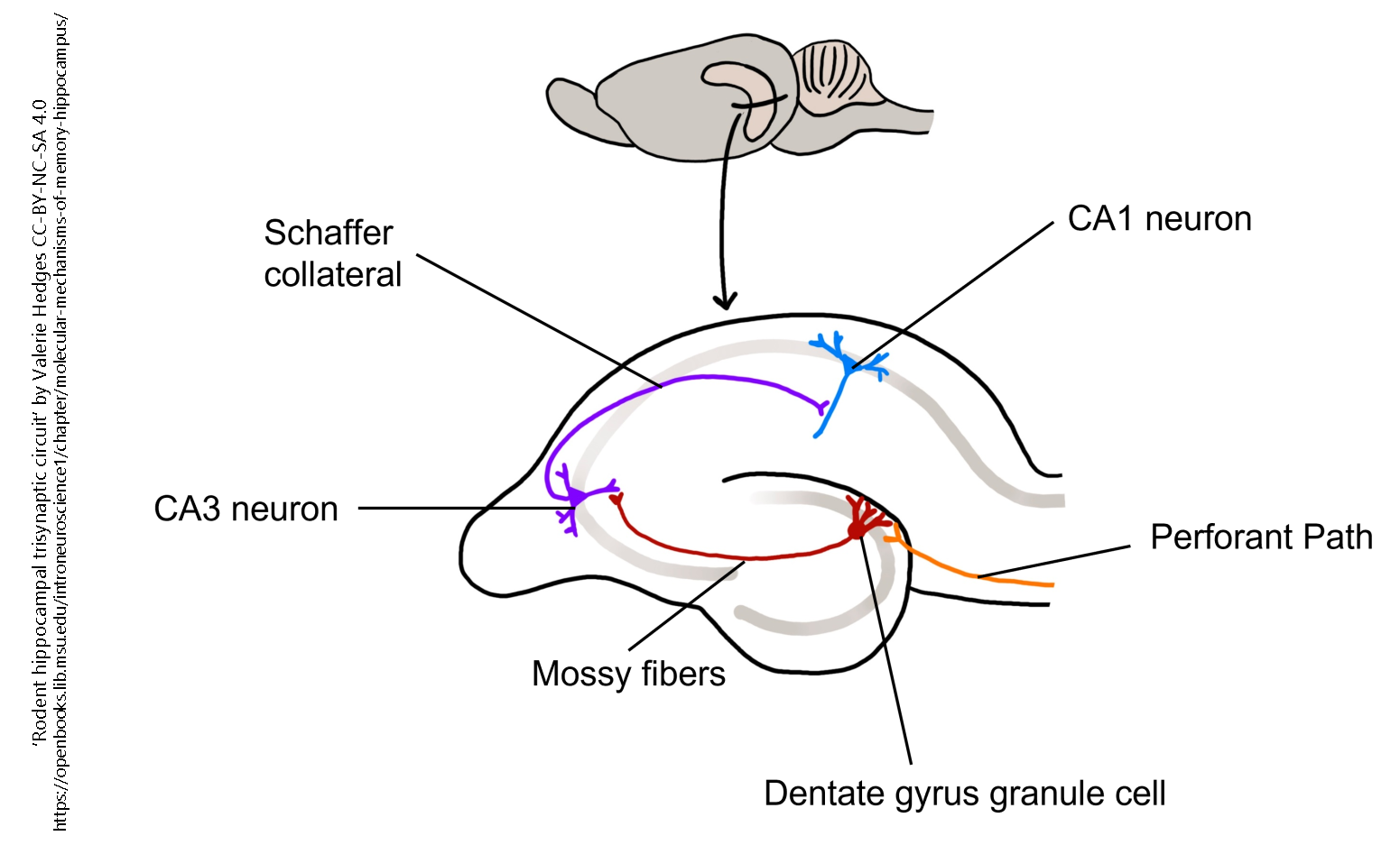
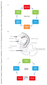
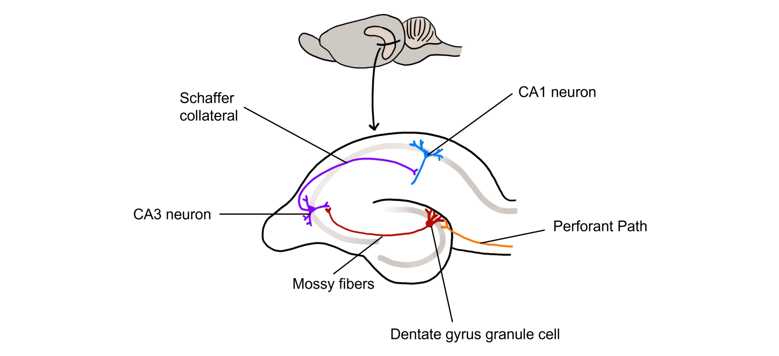
Over the years since, neuroscientists have focused on three circuits found in the hippocampal slice.
- the perforant path: from the entorhinal cortex to granule cells in the dentate gyrus;
- the mossy fiber pathway: the axons of dentate gyrus granule cells making synapses on the dendrites of CA3 neurons;
- the Schaffer collateral pathway: the axons of CA3 cells making synapses on the dendrites of CA1 neurons.
The perforant path was the circuit studied by Bliss and Lømo, and therefore the site of the first demonstration of a Hebbian synapse. More recently, most researchers study the Schaffer collaterals.
Media Attributions
- Hippocampus and seahorse © Laszlo Seress is licensed under a CC BY-SA (Attribution ShareAlike) license
- Hippocampus © Anatomography (Life Science Databases) is licensed under a CC BY-SA (Attribution ShareAlike) license
- Circuitry of the hippocampus © Pradip Chauhan, Kinjal Jethwa, Ashish Rathawa, Girish Chauhan, and Simmi Mehra is licensed under a CC BY-NC (Attribution NonCommercial) license
- Bliss and Lomo 1973 is licensed under a All Rights Reserved license
- Hippocampus Anatomy Hedges © Valerie Hedges is licensed under a CC BY-NC-ND (Attribution NonCommercial NoDerivatives) license
