The Endomembrane System
Jim Hutchins
Objective 4: Describe the endomembrane system and how it relates to the synthesis and release of neurotransmitters.
Neuronal Energy Objective 4 Video Lecture
The Endomembrane System

Early studies using electron microscopy seemed to indicate that the endoplasmic reticulum, the Golgi complex (Golgi apparatus), and vesicles were all separate entities. This is how they came to have separate names. However, more recent research shows that these are all part of a continuous network called the endomembrane system.
In this system, mRNA is brought from the nucleus, made into protein by the ribosomes of the rough endoplasmic reticulum. The newly-made (“nascent“) protein is transported to the cis face of the Golgi complex. After modification, it leaves the trans face of the Golgi and is contained a transport vesicle. (Note that we are using the terms cis and trans in their original organic chemistry sense; cis means on the same side and trans, on the opposite side, in this case relative to the rough endoplasmic reticulum.)
Some transport vesicles are used for intracellular processes, such as to destroy unwanted, unneeded, or malformed proteins by fusing them with lysosomes.
Some transport vesicles fuse with the plasma membrane (cell membrane) to insert pumps, channels, receptors, and other proteins in the membrane.
The transport vesicles which we’ll describe below are used to expel the proteins into the extracellular space, a process called exocytosis (literally, “process of taking something outside the cell“).
Exocytosis in Neurons
Exocytosis in neurons takes on a critical role because most neurons communicate with each other using chemical extracellular signals called neurotransmitters. Protein neurotransmitters follow the “standard” process of exocytosis as described for many other cells.
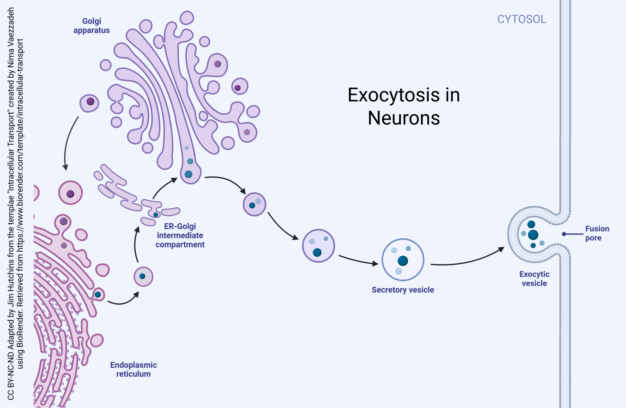
After protein is made, the process of protein modification begins.
First, proteins travel into the ER-Golgi intermediate compartment (ERGIC). The ERGIC vesicles fuse together to form the cis face of the Golgi. There are also retrograde transport vesicles which leave the cis face of the Golgi and return to the ER.
Anterograde transport vesicles move proteins (in a general cis-to-trans direction) between the stacks of Golgi membranes (cisternae). This process can be one or more of:
- protein refolding
- protein cleavage
- addition of chemical groups such as sugars or phosphate groups
- formation of protein complexes (several separate protein chains combined into one “package”, as in neurotransmitter receptors)
The completed protein is now ready to be packaged into a transport vesicle. If the transport vesicle is destined to be fused with the cell membrane, it’s called a secretory vesicle.
In neurons, protein modification might continue while it is being transported to a distant site. For example, pro-opiomelanocortin is a protein made by the rough endoplasmic reticulum of some neurons, and it is cleaved into several different bioactive compounds while it is being transported in a vesicle to the axon terminal.
Neurotransmitter Release Is a Specialized Kind of Exocytosis
 All cells are capable of secreting chemical substances to regulate the cells around them. The general name for this type of secretion is paracrine secretion to distinguish it from endocrine secretion, the release of chemical substances into the bloodstream to act on distant organs. Because neurotransmitters generally act over short distances (less than 1 μm) and for short periods of time, neurotransmission is a highly specialized and regulated form of paracrine secretion.
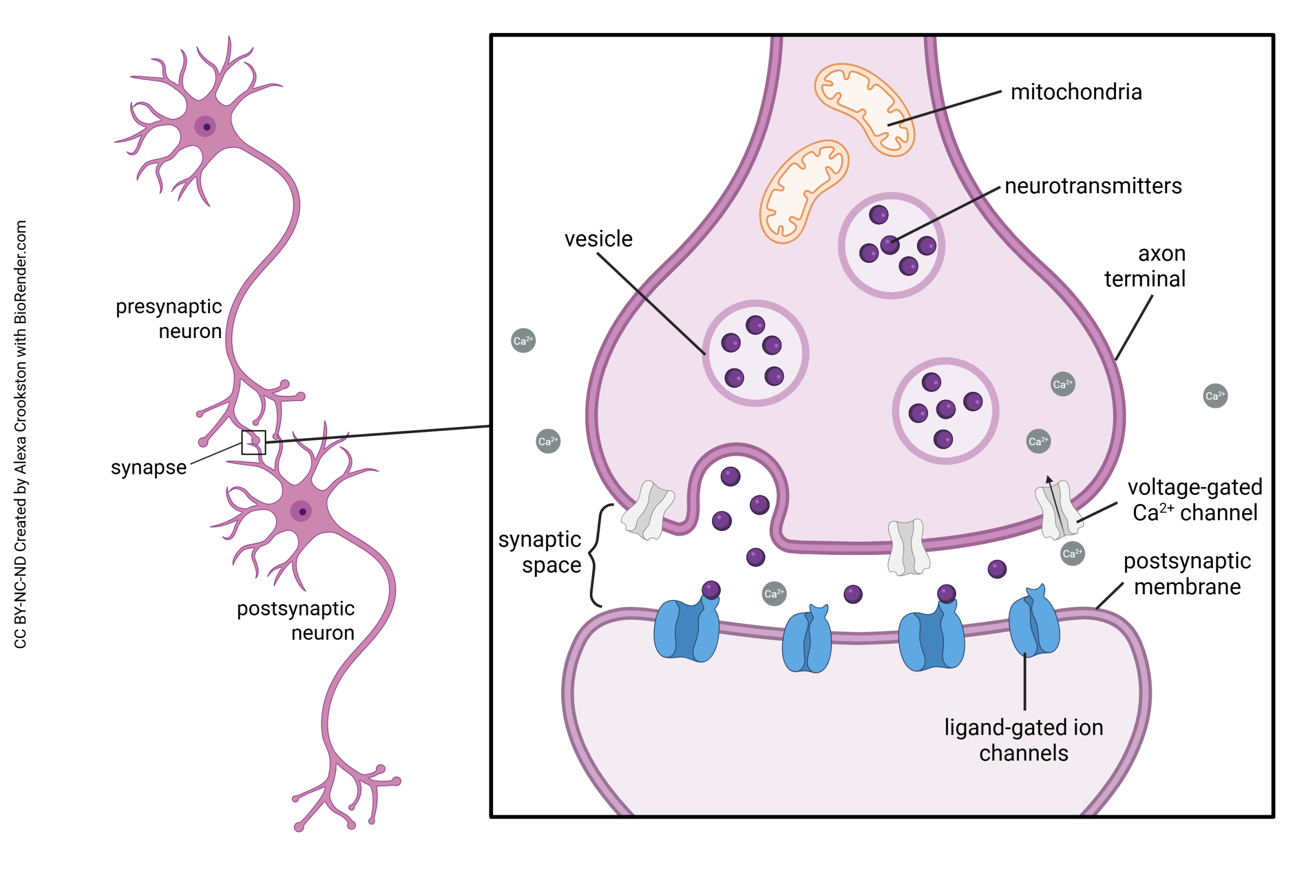
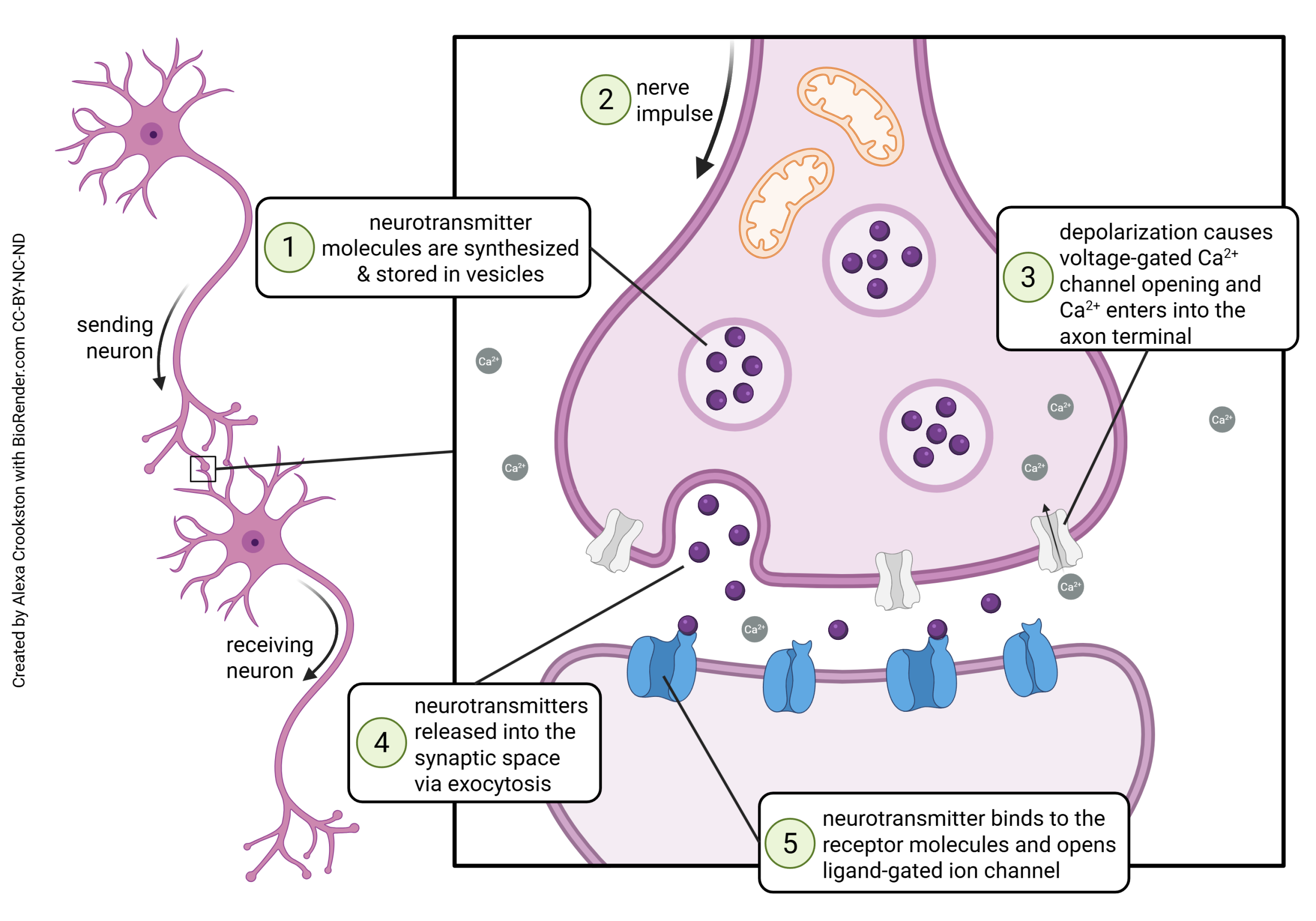 Neurotransmission occurs at a structure called the synapse. The word “synapse” (from the Greek word synaptein, “to clasp”) was coined by Charles Scott Sherrington in 1897 to describe a theoretical concept, long before anyone actually saw a synapse as drawn above.
Some of the steps in vesicle fusion and neurotransmitter release at the synapse will be discussed later. Now, we are focusing on the mechanisms of neurotransmitter exocytosis at the synapse.
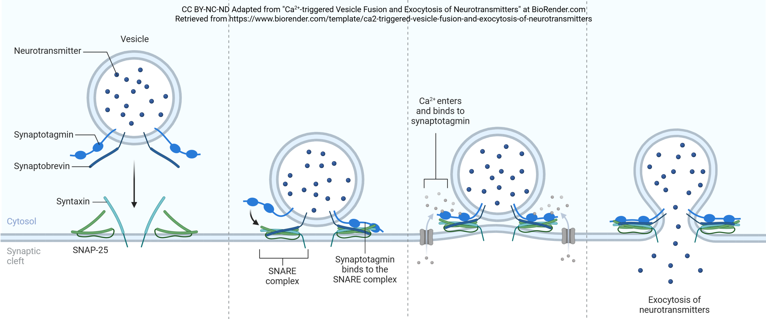 Synaptic vesicles are coated with a variety of proteins including synaptotagmin and synaptobrevin. Synaptobrevin is also called VAMP. The deadliest substance known to mankind, botulinum toxin, works by cleaving synaptobrevin and thereby blocking neurotransmitter release. (One gram of botulinum toxin, properly dispersed, would kill 1,000,000 people; gram for gram, it’s 1000X more deadly than plutonium.)
Synaptotagmin is sensitive to calcium ion (Ca2+) levels in the neuronal cytoplasm. When Ca2+ levels rise, synaptotagmin and synaptobrevin attach to two proteins that are found on the inner surface of the cell membrane at the synapse, syntaxin and SNAP-25. Together, these proteins form a SNARE complex. Ca2+ entry causes the SNARE complex to collapse, drawing the vesicle close to the presynaptic membrane. Fusion between the vesicle membrane and presynaptic membrane occurs, and neurotransmitter is released through exocytosis.
Regulated and Constitutive Secretion in Neurons
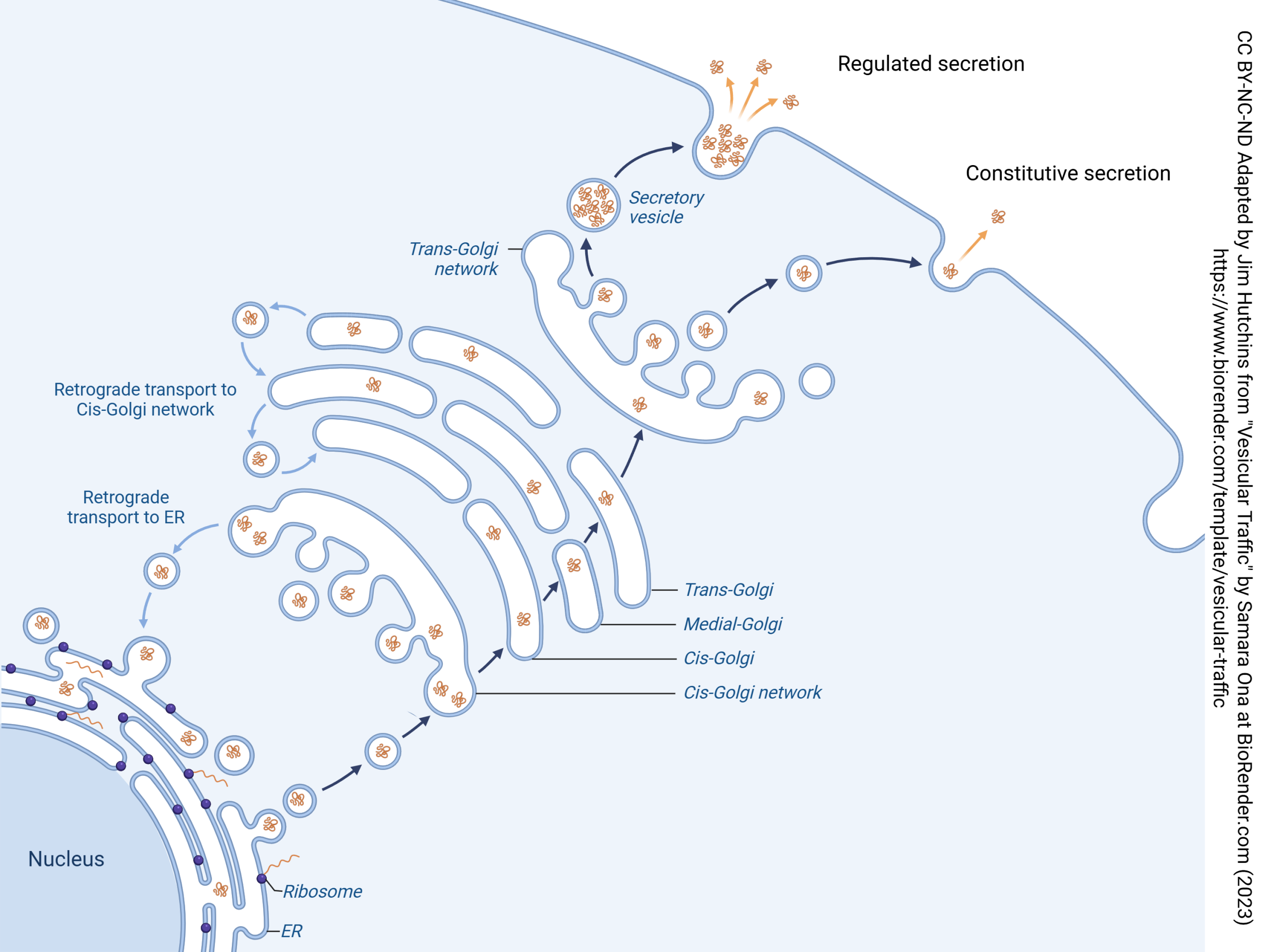
Vesicular release of neurotransmitter is an extreme example of regulated secretion: secretion that only occurs when specialized conditions exist. In the case of synaptic release, the specialized condition is entry of Ca2+. Neurons, and other cells, may regulate exocytosis through other signaling pathways.
Not all secretion is regulated. Neurons, and other cells, may constantly make proteins and release them 24/7/365.2422. This type of secretion is called constitutive secretion. The word “constitutive” is used by biologists to mean “occurring constantly”.
Endocytosis in Neurons
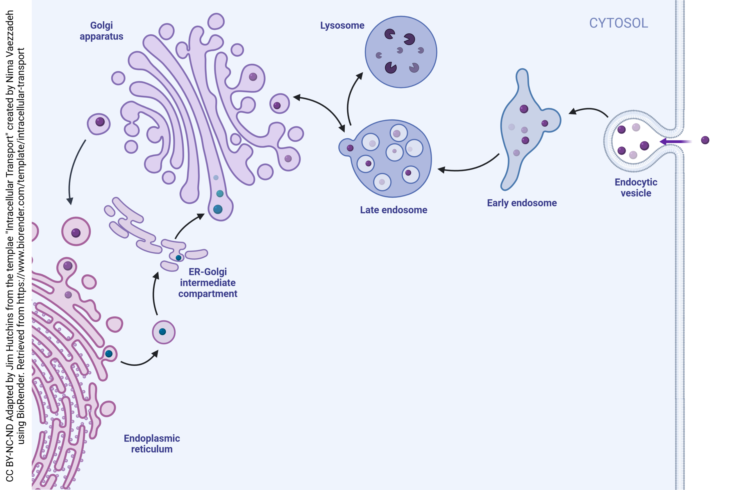
Neurons also take substances back into the cell and recycle vesicles. This process is called endocytosis. If there were no endocytosis, the surface area of the neuronal cell membrane would consistently increase in tiny increments until the cell became significantly larger. Also note that the vesicle membrane is made up of the same components as the cell membrane, which means that vesicular release and endocytosis do not change the markers found on the surface of the cell.
When substances are to be taken up by a neuron, they form an endocytic vesicle. Several endocytic vesicles fuse to form the early endosome. The early endosome has the same contents as the original endocytic vesicles.
Then, the late endosome forms through interaction with the trans face of the Golgi. Substances move back and forth between the late endosome and the Golgi. Eventually, the late endosome contents fuse with the lysosome and are destroyed. The constituent lipids, carbohydrates, and amino acids can then be recycled into other substances by the neuron.
Media Attributions
- Endomembrane system © Betts, J. Gordon; Young, Kelly A.; Wise, James A.; Johnson, Eddie; Poe, Brandon; Kruse, Dean H. Korol, Oksana; Johnson, Jody E.; Womble, Mark & DeSaix, Peter is licensed under a CC BY (Attribution) license
- Exocytosis in neurons © Nima Vaezzadeh adapted by Jim Hutchins is licensed under a CC BY-NC-ND (Attribution NonCommercial NoDerivatives) license
- Neurotransmitter System (anatomy) © Alexa Crookston is licensed under a CC BY-NC-ND (Attribution NonCommercial NoDerivatives) license
- Neurotransmitter System (action) © Alexa Crookston is licensed under a CC BY-NC-ND (Attribution NonCommercial NoDerivatives) license
- Ca2+-triggered Vesicle Fusion and Exocytosis of Neurotransmitters © BioRender adapted by Jim Hutchins is licensed under a CC BY-NC-ND (Attribution NonCommercial NoDerivatives) license
- Regulated and constitutive secretion © Samara Ona adapted by Jim Hutchins is licensed under a CC BY-NC-ND (Attribution NonCommercial NoDerivatives) license
- Endocytosis in Neurons © Nima Vaezzadeh adapted by Jim Hutchins is licensed under a CC BY-NC-ND (Attribution NonCommercial NoDerivatives) license
