The Cranial Nerves
Jim Hutchins
Objective 9: Label the 12 cranial nerves.
 The spinal cord has spinal nerves. These are segmental. The simplest explanation of “segmental” is that they cover a specific region of skin and/or muscle and are always mixed (sensory + motor).
Inside the skull, there is a different system. The presence of head-only muscle groups (e.g. for eye movements), head-only sensations (e.g. vision, hearing) and head-only autonomic functions (e.g. salivation) along with all the “regular stuff” means that the head needs cranial nerves.
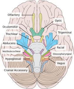
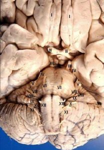
Classically, there are 12 cranial nerves. Twelve of anything is hard to remember, so students have had to spend lots of time coming up with silly mnemonics to recall these. We will give you a clean mnemonic here. Your instructor may share with you an off-color mnemonic if you ask. Or, you can look them up. Anything that involves Oliver is especially disgusting, so be careful what you look for.
 I. Olfactory
II. Optic
III. Oculomotor
IV. Trochlear
V. Trigeminal
VI. Abducens
VII. Facial
VIII. Vestibulocochlear
IX. Glossopharyngeal
X. Vagus
XI. Accessory (Spinal Accessory)
XII. Hypoglossal
You should become skilled at naming these using both the Roman numeral designation and the full name, and know a little bit about the anatomy and function of each. We’ll go through them one at a time.
The clean mnemonic (using the first letter of the name of each cranial nerve) is:
“Oh, Oh, Oh, To Touch And Feel Very Green Vegetables, AH!”
To remember which are Sensory, which are Motor, and which are Both: Some Say Marry Money, But My Brother Says Big Brains Matter More
The first two cranial nerves are really just central nervous system tracts. Both carry sensory information: olfactory (Latin olfacere, “to smell”) and optic (Greek οπτικος, optikos, “visible”).
Cranial Nerve I: Olfactory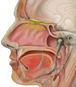
The olfactory nerve carries olfactory (i.e. smell) information from the olfactory epithelium inside the nasal sinuses to the brain.
Cranial Nerve II: Optic
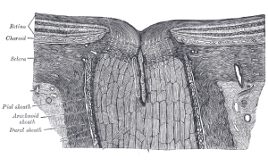
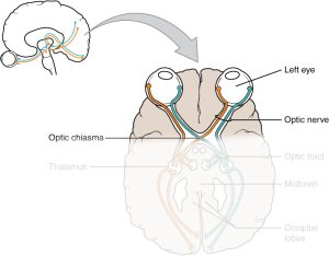
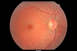
The optic nerve carries optic (i.e. sight, visual) information from the retina lining the inside of the eye to the brain.
Cranial Nerve III: Oculomotor
Cranial Nerve IV: Trochlear
Cranial Nerve VI: Abducens
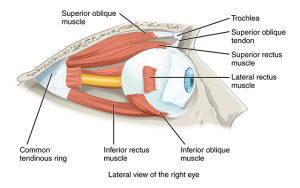
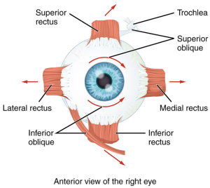 The next three cranial nerves innervate the extraocular muscles. These muscles move the eye around in the orbit, so each nerve is purely motor.
The oculomotor nerve innervates four of the six muscles: medial rectus, superior rectus, inferior rectus, and inferior oblique muscles. It also innervates the iris muscles which control the size of the pupil.
The trochlear nerve innervates the superior oblique muscle. The superior oblique passes through a bony loop in the superior part of the orbit. This bony loop is called the trochlea (Latin, “pulley”).
The abducens nerve is so named because it abducts the eye (carries it away from the midline), so it innervates the lateral rectus.
Cranial nerves V and VII innervate the face. Both are mixed (sensory + motor) nerves, but V is primarily sensory for the face while VII is primarily motor for the muscles of facial expression.
Cranial Nerve V: Trigeminal
The trigeminal nerve is named “tri-” because it has three divisions: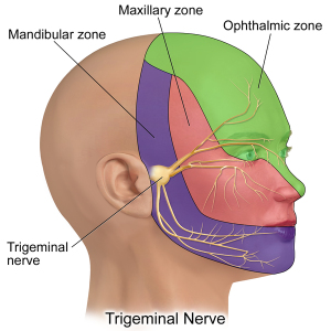
- V1 or ophthalmic for the eyes and forehead;
- V2 or maxillary for the upper jaw region (maxilla);
- V3 or mandibular for the lower jaw region (mandible).
The trigeminal nerve is the largest cranial nerve.
Management of the trigeminal nerve is a critical part of dental practice. Trigeminal pain (such as from a tooth abscess) is excruciating and patients will suffer until it is relieved.
The trigeminal nerve also has the sensory function of proprioception for the jaw muscles. This keeps us from crushing our teeth by biting down too hard on a nut or seed: the trigeminal helps the jaw muscles relax as soon as the force on the teeth releases.
The motor function of the trigeminal nerve is for the muscles of mastication. These are the four chewing muscles, and four more, which we will not name here.3
Cranial Nerve VII: Facial
The facial nerve (cranial nerve VII) is primarily motor for the muscles of facial expression, but has some other motor functions and sensory function as well.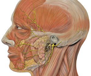
In addition, it innervates the lacrimal glands (Latin lacrimare, “to weep”) which produce tears and the submandibular and sublingual glands which produce saliva.
The sensory function of the facial nerve is to carry taste information from the anterior 2/3 of the tongue. This information joins with the other taste fibers to form a special nerve called the chorda tympani which is not one of the cranial nerves but important to know.
3 OK, fine. Masseter, temporalis, medial pterygoid, lateral pterygoid, tensor tympani, tensor veli palatini, myelo-hyoid, and the anterior belly of the digastric. Happy now?
Cranial Nerve VIII: Vestibulocochlear
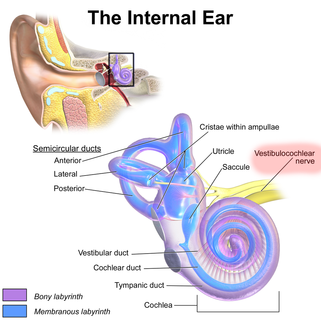
The eighth cranial nerve carries sensory information from the special sense organs of the inner ear.
The vestibulocochlear nerve, as the name tells us, has two divisions. On the gross view of the brain, the divisions can easily be seen; the nerve looks like a figure 8 in cross-section.
The cochlear branch of CN VIII carries information from the spiral-shaped cochlea (Latin: “snail”). The cochlea is responsible for taking sound waves from the environment and coding the information in a form the nervous system can use.
The vestibular branch of CN VIII carries information from the semicircular canals and vestibule. The semicircular canals code for head acceleration (rotation) in each of the three major planes. The vestibule (saccule and utricle) codes for head movement. The saccule and utricle are also called the otolith organs (Greek: οτο, oto, “ear”; λιθος, lithos, “stone”).
Cranial Nerve IX: Glossopharyngeal
Cranial Nerve X: Vagus
Cranial Nerve XII: Hypoglossal
The glossopharyngeal nerve innervates the posterior 1/3 of the tongue (Greek, γλωσσα, glossa, “tongue”) and the pharynx.
Remember that taste information for the anterior 2/3 of the tongue is carried on the facial nerve. Taste for the remainder of the tongue is carried on CN IX.
CN IX also innervates the parotid gland, the largest of the salivary glands.
Another important homeostatic function of the glossopharyngeal nerve is to carry information from the carotid body, a sensory organ on the carotid artery which senses blood pressure.
The vagus nerve (Latin, “wandering”) travels widely over the neck, thorax and abdomen, innervating a wide variety of internal organs. If someone’s words wander all over the place, we call them “vague.” If a nerve wanders all over the place, we call it the vagus. It carries some taste information from taste buds in the soft palate and uvula (the “hangy thing” at the back of your throat, sorry for the technical language). Mostly, however, the vagus carries autonomic parasympathetic information to the internal organs.
The hypoglossal nerve (Greek, υπο–, ypo–, “under” plus –γλωσσα, –glossa, “tongue”) is purely motor, and does only one thing (but it does it quite well): it sticks out the tongue. One odd thing is that the tongue is pulled forward out of the mouth, since muscles can only pull and not push.
Cranial Nerve XI: (Spinal) Accessory Nerve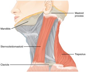
The accessory nerve (also called the spinal accessory nerve) is pure motor, innervating two muscles: the trapezius along the back of the neck and shoulders, and the sternocleidomastoid, which connects the sternum (sterno–), clavicle (–cleido–), and – mastoid process of the temporal bone.
Media Attributions
- Cranial nerves human brain normal inferior view © Lynch, Patrick J. adapted by Jim Hutchins is licensed under a CC BY-SA (Attribution ShareAlike) license
- Human brain anterior inferior view © Beal, John A. PhD adapted by Jim Hutchins is licensed under a CC BY (Attribution) license
- Primary Terminal © Gray, Henry is licensed under a Public Domain license
- Nucleus of Origin © Gray, Henry is licensed under a Public Domain license
- Olfactory nerve © Lynch, Patrick J. is licensed under a CC BY (Attribution) license
- Optic nerve head longitudinal © Gray, Henry is licensed under a Public Domain license
- Optic Nerve © Betts, J. Gordon; Young, Kelly A.; Wise, James A.; Johnson, Eddie; Poe, Brandon; Kruse, Dean H. Korol, Oksana; Johnson, Jody E.; Womble, Mark & DeSaix, Peter is licensed under a CC BY (Attribution) license
- Human retina fundus © Manuel Pinot is licensed under a CC BY-NC-ND (Attribution NonCommercial NoDerivatives) license
- Extraocular muscles © Betts, J. Gordon; Young, Kelly A.; Wise, James A.; Johnson, Eddie; Poe, Brandon; Kruse, Dean H. Korol, Oksana; Johnson, Jody E.; Womble, Mark & DeSaix, Peter is licensed under a CC BY (Attribution) license
- Extraocular muscles © Betts, J. Gordon; Young, Kelly A.; Wise, James A.; Johnson, Eddie; Poe, Brandon; Kruse, Dean H. Korol, Oksana; Johnson, Jody E.; Womble, Mark & DeSaix, Peter is licensed under a CC BY (Attribution) license
- Trigeminal Nerve © BruceBlaus is licensed under a CC BY-SA (Attribution ShareAlike) license
- Facial nerve branches © Lynch, Patrick J. is licensed under a CC BY (Attribution) license
- Vestibulocochlear Nerve Ear Anatomy © BruceBlaus is licensed under a CC BY (Attribution) license
- Glossopharyngeal vagus hypoglossal © Gray, Henry is licensed under a Public Domain license
- Accessory nerve muscles © Betts, J. Gordon; Young, Kelly A.; Wise, James A.; Johnson, Eddie; Poe, Brandon; Kruse, Dean H. Korol, Oksana; Johnson, Jody E.; Womble, Mark & DeSaix, Peter is licensed under a CC BY (Attribution) license
