The Convergence of Advances in Physiology, Histology, and Electricity Lead to an Acceleration in Our Understanding of the Brain
Jim Hutchins
Objective 3: Relate how the study of physiology, histology, and electricity led to a revolution in the understanding of neuroscience.
History of Neuroscience Objective 3 Video Lecture

At the beginning of the 19th century, neuroscience was a very tiny corner of biology. Any field of biology that existed in the 19th century, however, was greatly advanced by the observations of Charles Darwin as he voyaged on the HMS Beagle from 1831 to 1836. After becoming famous for his diary of the voyage, Darwin reorganized his observations on natural history as The Origin of Species, published in 1859. In it, Darwin proposed a theory of natural selection. There are two key elements to the theory. Traits are inherited from the parent, and vary amongst members of a species. Traits which increase fitness (i.e., the ability to reproduce) are more likely to be passed on to offspring; traits which decrease fitness are less likely to be passed on.
Behavior is a heritable trait, just like any other. This allows us to study the brain of different species to determine how those species are adapted to their individual ecological niches.
For example, dogs have an area in the frontal cortex called the prorean (or proreal) gyrus. Much of the herding behavior of the dog is due to activity of brain cells in the prorean gyrus. A dog’s primordial ancestors had a rudimentary prorean gyrus which these species used to hunt in packs. Humans found this behavior useful for hunting, and over 30,000 years, selectively bred dogs for exceptional herding ability. At the end of this process, the prorean gyrus has become a much more elaborate and well-developed structure. This structural change parallels the incredible increase in herding ability in the modern domestic dog. The inheritance of a behavior (herding ability) is reflected in an anatomical change over evolutionary time.
As we select for behaviors in breeding experiments, we change the wiring and therefore the anatomy of the brain.
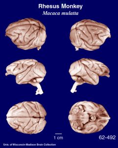 Monkeys must find food by color and detail. The monkey visual cortex, in the back of the brain (toward the center of these photographs), is a highly organized and elaborate brain structure.
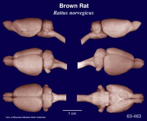 Rats crawl around in the dark. Rats don’t need much visual cortex (in fact, in the lab, albino rats are essentially blind, yet they appear to behave pretty much like brown sighted rats). They do need detailed sensation from their vibrissae, the whiskers that protrude from their muzzle. In the brain cortex which senses touch, there are elaborate structures called barrel fields whose organization reflects this important function.
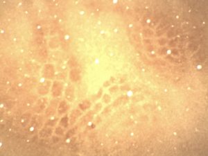
This photomicrograph of a flattened rat brain is stained for the mitochondrial enzyme cytochrome oxidase.
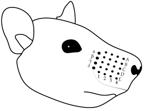
Each dark spot (barrel) represents exactly one vibrissae (hair on the muzzle).
Along with the advances in neuroscience driven by our understanding of natural selection, our advancement in an understanding of the electrical basis for nerve conduction came with another set of questions (as advances always do).
We know that electrical potentials spread in both directions along a wire. But is that true of electrical potentials traveling in the nervous system? The way in which nerve information travels as electrical potentials in the nervous system is governed by the Bell-Magendie Law.
About 1810, Charles Bell (working in Scotland) and François Magendie (working in France) more-or-less simultaneously approached the same question in the same way: are nerves unidirectional, sending information in one direction, or bidirectional like a wire, sending information in both directions?
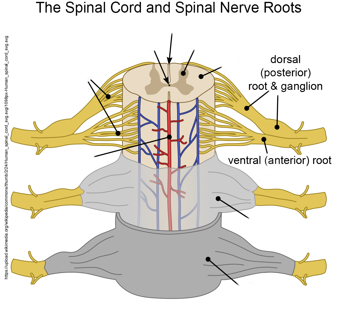 The spinal cord has dorsal (posterior) and ventral (anterior) roots, as shown in the diagram. (Dorsal means “toward the back” and ventral means “toward the belly”; the terms posterior “back” and anterior “front” replace these in human anatomy.)
If Bell or Magendie cut the ventral (anterior) roots, paralysis resulted. The animal was unable to move a limb on the operated side.
If they cut the dorsal (posterior) roots, loss of sensation resulted.
Two important observations resulted from these experiments:
- the dorsal part of the spinal cord is responsible for managing sensory (feeling) information, while the the ventral part is responsible for generating motor (movement) information;
- individual nerve fibers carry information in only one direction under physiological conditions.
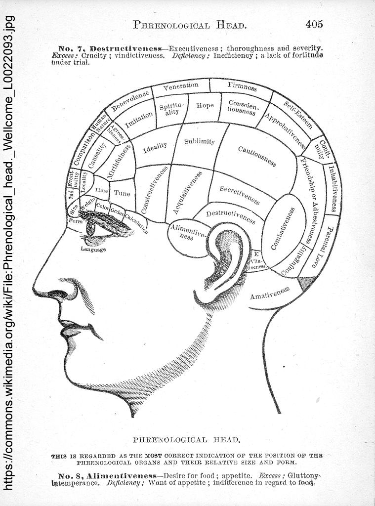 Just as the dispute between Galvani and Volta dominated the late 18th century discussion of the electrical properties of the nervous system, and advanced the field in doing so, there was a less-polite scientific disagreement between Franz Joseph Gall and Marie Jean Pierre Flourens. Gall’s ideas came to predominate 19th century neuroscience. He believed (correctly) that most brain functions were localized to specific areas of the human brain. He then concluded (incorrectly) that bumps on the skull were correlated with areas of larger and more complex development. Gall was responsible for the pseudoscience of phrenology, where a “practitioner” would feel the bumps on the skull and make representations about a person’s abilities and interests.
Flourens, who believed (correctly) that the cerebellum is involved in the control of movement, criticized Gall on three grounds:
- ablation of the brain areas identified by Gall does not produce the results Gall would predict (correct);
- just because a bump exists on the skull, doesn’t mean it has anything to do with the structure or size of the underlying brain part (correct);
- all areas of the cerebrum participate equally in sensory and motor functions (wrong).
Media Attributions
- HMS Beagle © John Chancellor & Karen E James is licensed under a CC BY-NC-SA (Attribution NonCommercial ShareAlike) license
- Rhesus monkey brain © University of Wisconsin-Madison Brain Collection is licensed under a Public Domain license
- Brown rat brain © University of Wisconsin-Madison Brain Collection is licensed under a Public Domain license
- Rat Barrel Cortex © Imbrickle is licensed under a Public Domain license
- Vibrissae sensors © Scharff, Moritz, Philipp Schorr, Tatiana Becker, Christian Resagk, Jorge H. Alencastre Miranda, and Carsten Behn adapted by Jim Hutchins is licensed under a CC BY (Attribution) license
- Human spinal cord and spinal nerve roots © Tomáš Kebert & umimeto.org adapted by Jim Hutchins is licensed under a CC BY-SA (Attribution ShareAlike) license
- Phrenological head © Wellcome Collection is licensed under a CC BY (Attribution) license

