The Midbrain
Jim Hutchins
Objective 6: Locate the major structures of the midbrain.
Important Locations Objective 6 Video Lecture
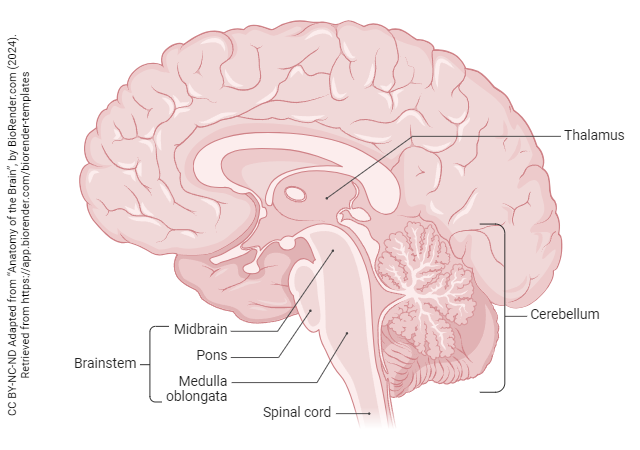
The brainstem is divided into three regions and a sort-of annex. From rostral (top) to caudal (bottom), they are the:
- midbrain
- pons
- medulla oblongata
The midbrain is the smallest major division of the brain. The most important function of the midbrain is controlling visual reflexes, like pupil size. When we look at a patient’s pupils with a flashlight, we are assessing the function of the midbrain.
Nuclei of the Midbrain Tegmentum
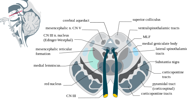
The midbrain is divided into a tegmentum (Latin, “covering”) and a tectum (Latin, “roof”). A pipe-shaped cerebral aqueduct connects the third ventricle and the fourth ventricle, reservoirs of cerebrospinal fluid.
The oculomotor nerve (cranial nerve III) controls four of the six extraocular muscles (medial rectus, superior rectus, inferior rectus, and inferior oblique). These muscles are skeletal (voluntary) muscle. Cell bodies which reside in the medial part of the midbrain, just anterior to the cerebral aqueduct, send axons via CN III to make neuromuscular junctions on these muscles.
Nestled in the groove of the V-shaped oculomotor nucleus is a subdivision of CN III called the Edinger-Westphal nucleus. These cell bodies will innervate the smooth muscle of the pupillary sphincter to reduce pupil size when they are activated, a parasympathetic response. The same nucleus contains cell bodies of neurons which control lens shape and focal length. These are all autonomic responses, so the Edinger-Westphal nucleus is the autonomic division of the nucleus (collection of cell bodies) of the third cranial nerve.
The most prominent feature, and largest nucleus, of the midbrain tegmentum is the substantia nigra (Latin, “black substance”). These cells primarily produce the neurotransmitter dopamine and, as a by-product, the pigment melanin which gives this area a black or dark brown color. Dopamine produced in the substantia nigra is released in the basal nuclei to help control movement. The loss of these critical cells leads to Parkinson’s disease.
The trigeminal nerve (cranial nerve V) is the major source of sensory input from the face. Cell bodies which will sense the position of the jaw (proprioception) are found in the mesencephalic nucleus of V.
The red nucleus is also found in the midbrain tegmentum. This large, prominent area with a rich supply of iron (hence the red color, and its name) is not well-studied but is believed to be involved in the control of the upper extremities.
There is a loose network of cells and fibers called the reticular formation at all levels of the brainstem; the part of this complex which resides in the midbrain is the midbrain reticular formation.
The Midbrain Tectum
The midbrain tectum is generally called, simply, “the tectum”. It consist of four symmetrical bumps collectively called the corpora quadrigemina (Latin, “four twinned bodies”). These are the right and left superior colliculus and the right and left inferior colliculus. Colliculus, in Latin, means “little hill”. The superior colliculus or optic tectum has been extensively studied, especially its role in the unconscious processing of visual information. It is thought to be the center responsible for a surprising phenomenon called blindsight in humans: the ability to avoid objects in one’s path, even in the absence of conscious vision.
Just as the superior colliculus makes the calculations needed for visual reflexes, the inferior colliculus performs calculations needed to support auditory reflexes.
Major Fiber Pathways Passing Through the Midbrain
Axons which originate in the primary motor cortex (Brodmann 4) travel in the crus cerebri, a large bump on the anterior surface of the brainstem. If one focuses on the structures which connect the midbrain to the cortex, then these may be called the cerebral peduncles (Latin: “little foot of the brain”). Functionally, these axons are called the corticospinal tract. The axons of the corticospinal tract are arranged by the muscles they will eventually innervate, by synapsing on cell bodies of alpha motor neurons in the spinal cord. Axons controlling the face are most medial. Then, from medial to lateral, we find fibers controlling the arm (upper extremity); trunk; and lower extremity. The mnemonic for this pattern is FATL. The corticospinal tract makes up about the middle 3/5 of the crus cerebri.
Also in the midbrain are axons relaying sensory information from skin and muscle to the postcentral gyrus (primary somatosensory cortex), where it reaches conscious perception. Pain and temperature information is carried on the anterolateral system, which some older authorities call the spinothalamic tract. While it’s true that the anterolateral system relays information from the body to the thalamus, the information is also sent to many other functionally important locations, including the gray matter surrounding the cerebral aqueduct (the periaqueductal gray) and the raphé (French: “seam”) nuclei. The periaqueductal gray is a rich source of endogenous opioids like endorphins and enkephalins, while the raphé nuclei release serotonin. Both of these neurotransmitters are released into the posterior (dorsal) horn of the spinal cord, near the sensory receptor synapses onto relay cells, where they can modify or block incoming pain signals.
Proprioception, the ability to determine if you are being touched once or twice when adjacent skin areas are touched (“two-point discrimination”), light touch, and vibration, are all carried in the medial lemniscus. This type of information enters the posterior (dorsal) horn and synapses for the first time in the medulla; the medial lemniscus has axons that are the second neuron to carry this information.
Media Attributions
- Anatomy of the Brain © BioRender adapted by Jim Hutchins is licensed under a CC BY-NC-ND (Attribution NonCommercial NoDerivatives) license
- Mid-brain cross section © Madhero88 is licensed under a CC BY-SA (Attribution ShareAlike) license
