Targets of the Retinofugal Projection: Lateral Geniculate Nucleus, Midbrain, and Hypothalamus
Jim Hutchins
Objective 10: Describe the retinofugal pathway to the lateral geniculate nucleus, midbrain, and hypothalamic nuclei.
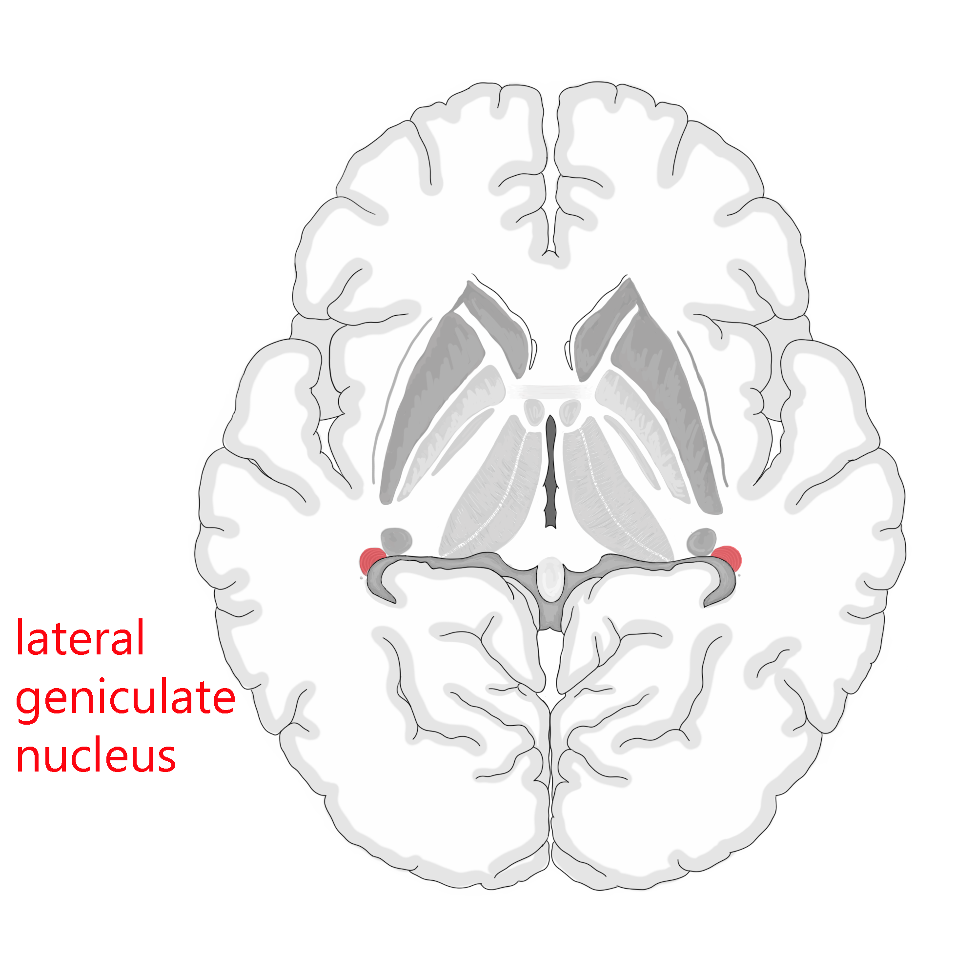
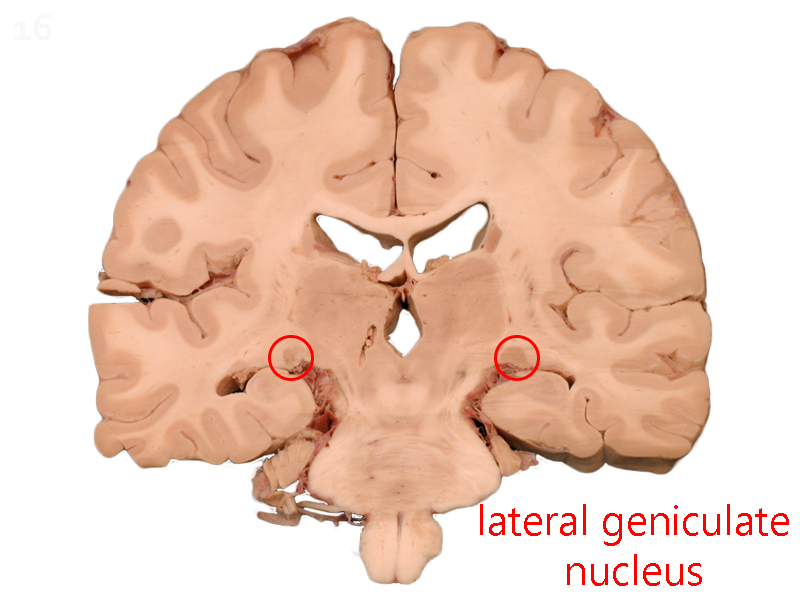
The optic tract ends in a structure called the lateral geniculate nucleus (LGN). The Latin term genu means “knee”, so “geniculate” means “bent like a knee”.
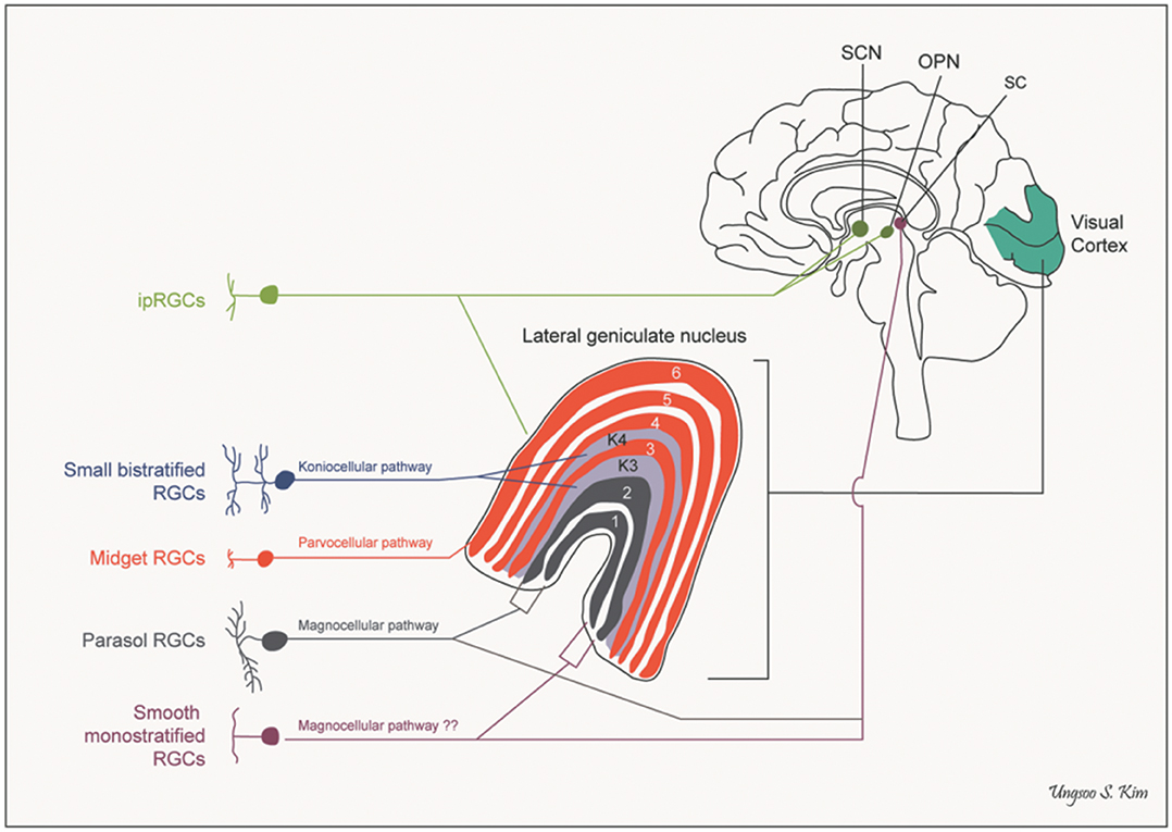
There are six layers of cells in the human LGN. Two (termed, naturally, layers 1 and 2) contain large cells and are called the magnocellular layers. Four (layers 3, 4, 5, and 6) contain smaller cells and are called the parvocellular layers. Late in our study of the LGN, we found that much of the action was occurring in even smaller cells in the LGN scattered throughout and between the layers but we had run out of names for sizes (magno– = “large”; parvo– = “small”) so in desperation, we called those koniocellular cells from the Greek konio– = “dust”.
Layers 1, 4, and 6 are innervated by ganglion cell axons from the contralateral side. Layers 2, 3, and 5 are innervated by ganglion cell axons from the ipsilateral side. M ganglion cells terminate in the magnocellular layers and P ganglion cells terminate in the parvocellular layers. (Non-M, non-P ganglion cells terminate onto the koniocellular cells.) So, in summary,
- Magnocellular layers
- 1: contralateral M parasol ganglion cells
- 2: ipsilateral M parasol ganglion cells
- Parvocellular layers
- 3: ipsilateral P midget ganglion cells
- 4: contralateral P midget ganglion cells
- 5: ipsilateral P midget ganglion cells
- 6: contralateral P midget ganglion cells
Each layer, then, contains a complete representation of objects in the contralateral visual field, with the same point in visual space “stacked” on top of a cell that conveys information about the same point. Like a toothpick in a layer cake, the same point in visual space is represented six times in the six layers of the LGN.
Superior Colliculus (Optic Tectum)
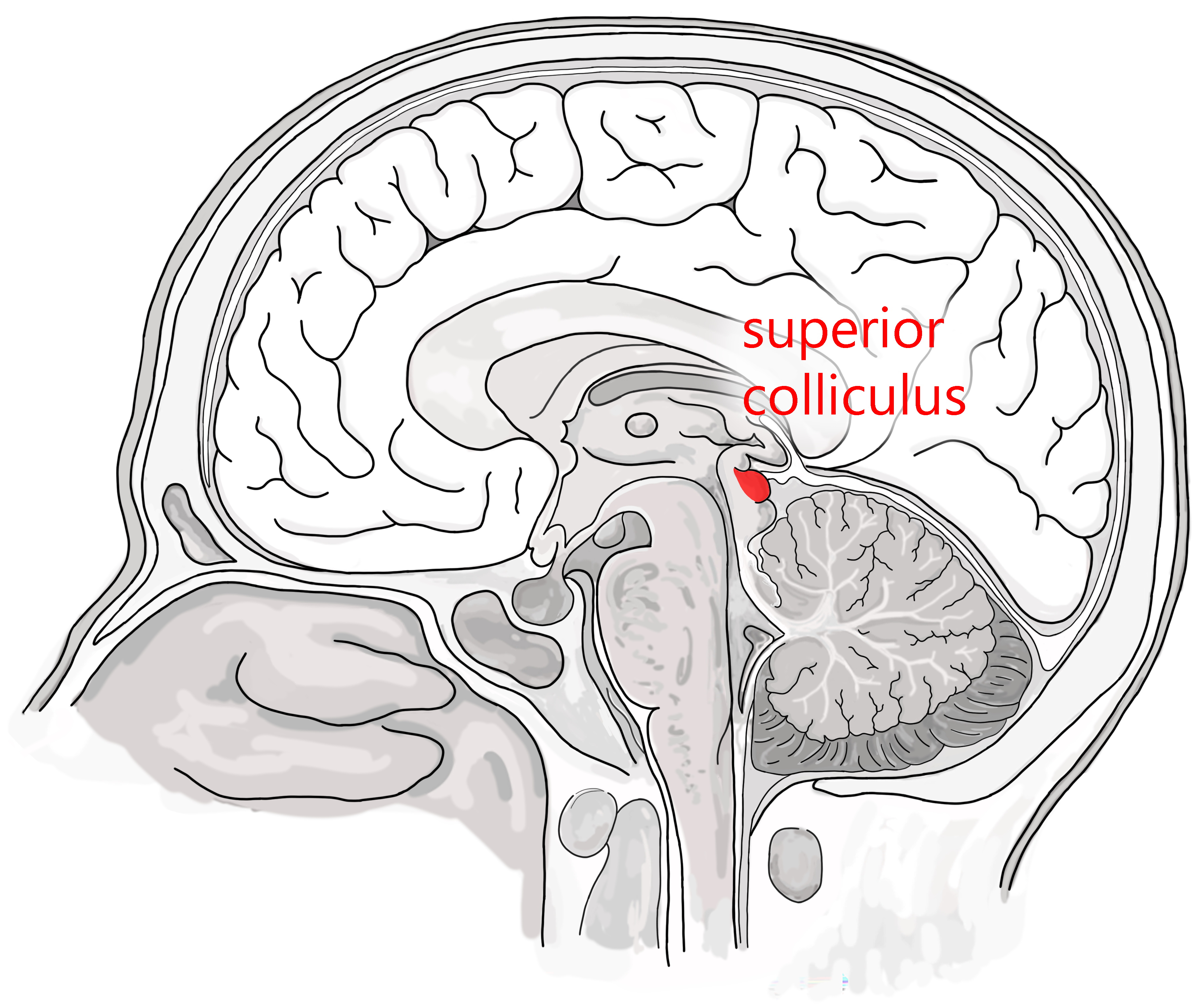
In the primate visual system, about 1 in 20 axons leaving the retina send a branch to the superior colliculus, a midbrain structure that mediates visual reflexes but does not participate in the conscious perception of visual stimuli.
The superior colliculi are a pair of bumps on the dorsal surface of the brainstem, collectively called the tectum. The Latin word tectum means “roof”, and these are named because they form the roof of the midbrain. In this naming system, the superior colliculus is called the optic tectum.
The optic tectum is the entire visual system of most vertebrates, since they have no cortex. For example, studies of how retinal cell axons form a specific pattern in the frog or fish visual systems looks at the optic tectum. These studies are an important part of developmental neuroscience.
Because we have no conscious perception of the activity of the superior colliculus, damage to the visual cortex which occurs (rarely) without damage to the retinotectal projection can cause a phenomenon called blindsight, where a person who is perceptually blind can still avoid objects placed in their path. This is illustrated in the following video.
Brainstem Nuclei Controlling Pupil Size
Because the eye must control the amount of light reaching the retina, so that it is roughly equal in bright sunlight or dim interior rooms, the midbrain nuclei controlling pupil size must receive some input from the retinal ganglion cells which transmit information about overall light levels. Recall that these melanopsin intrinsically photosensitive ganglion cells have receptive fields that cover the entire retina. Axons of these large-receptive-field ganglion cells project first to the pretectal olivary nucleus (PON) of the midbrain, and the PON in turn sends short axons to the nearby Edinger-Westphal nucleus, the autonomic motor nucleus which controls pupil size.
Optic Nerve Projections to the Hypothalamus
 A small minority of the retinal ganglion cell axons of the optic nerve project to the hypothalamus. The main hypothalamic target of these axons is the suprachiasmatic nucleus (SCN), named this because it is just dorsal (superior) to the optic chiasm.
The SCN is the primary region responsible for circadian rhythms, the daily cycles of activity that are common to all mammals.
Media Attributions
- Lateral geniculate nucleus © Avalon Marker and Jim Hutchins is licensed under a CC BY-SA (Attribution ShareAlike) license
- Coronal section of brain showing lgn © Functional Neuroanatomy adapted by Jim Hutchins is licensed under a CC BY-SA (Attribution ShareAlike) license
- Ganglion cells wired to lgn © Ungsoo Samuel Kim is licensed under a CC BY (Attribution) license
- Superior colliculus © Avalon Marker and Jim Hutchins is licensed under a CC BY-SA (Attribution ShareAlike) license
- Hypothalamus all nuclei © Avalon Marker and Jim Hutchins is licensed under a CC BY-SA (Attribution ShareAlike) license
