Organization of the Somatic Motor System
Caleb Bevan
Objective 1: Interpret a flowchart showing the organization of the somatic nervous system.
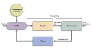
One common way to look at the function of systems is as a homeostatic loop. This is a perfect way to organize our thinking about motor systems: a system consists of input from receptors, processing by a set of neurons who extract information from the input and organize it in various ways; and an output to effectors.
Motor Systems Are the Effectors
 The nervous system as a whole contains probably dozens of levels of homeostatic loops all nested within each other, or in computer programming terms, subroutines that carry out different tasks.
Motor systems are all the parts of the brain and spinal cord that are devoted to the output of the nervous system. Recall that the nervous system has sensory, processing, and motor functions. The motor functions are the only outputs of the nervous system.
Divisions of the Motor Systems
There are two kinds of motor systems:
- autonomic (involuntary)
- somatic (voluntary)
We covered the autonomic motor system in a previous chapter. The embryonic structures that develop into voluntary (skeletal) muscle are the effectors of the somatic motor system. This objective is about the neural circuitry that controls these skeletal muscle effectors.
The embryonic structures that develop into smooth muscle, cardiac muscle, and glands are collectively called the visceral motor components by neuroembryologists and the autonomic nervous system and enteric nervous system by neurophysiologists. For example, the muscles that control the diameter of blood vessels, and therefore your blood pressure, are part of the autonomic nervous system. Similarly, the system that controls the rate at which your heart contracts is also part of the autonomic nervous system. The salivary gland secretions are part of this autonomic nervous system. The movement of substances through the gut tube (stomach and intestines) is controlled by the enteric nervous system which is closely tied to the autonomic nervous system and sometimes included with it.
Here is the overall organization of the nervous system. Motor systems are red in this diagram.

Media Attributions
- Homeostasis © Open Learning Initiative is licensed under a CC BY-NC-SA (Attribution NonCommercial ShareAlike) license
- Receptors © Lizz Bizzell is licensed under a CC0 (Creative Commons Zero) license
- Effectors © Lizz Bizzell is licensed under a CC0 (Creative Commons Zero) license
- Organization of nervous system © Jim Hutchins is licensed under a CC BY-SA (Attribution ShareAlike) license
