Skeletal Muscle
Objective #: Statement of objective.
Almost half of an adult’s body weight comes from muscle, one of the four main types of tissues found in the body. Although there are three different types of muscle tissue, they all share some basic characteristics. Muscle tissue is often referred to as “excitable,” meaning the electrical state of the plasma membrane can change. This change, known as depolarization, occurs in response to signals from the nervous system, endocrine system or as a reaction to various intercellular signaling molecules.. Regardless of muscle tissue type, the end result of depolarization is a shortening of fibers resulting in muscle contraction.
Muscle cells, also known as muscle fibers, share many of the same basic structures and functions as other cells. They have nuclei, cell membranes, mitochondria, cytoskeleton, as well as the other organelles found in body cells. They perform metabolism, communicate with other cells and are prone to cell damage and death. The most significant difference between muscle cells and other cells of the body is the density of proteins found within and surrounding the muscle cell. These proteins perform essential functions needed to allow the cell to expand and contract, which is the basis for movement in the body.
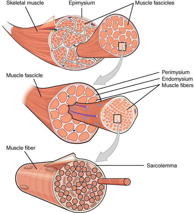
The most significant and most abundant constituents of muscle fibers are the contractile proteins actin and myosin. Simply put, myosin proteins pull on actin proteins, resulting in a shortening of muscle fibers, which leads to muscle contraction. The arrangement and specific mechanisms of action between actin and myosin varies between the different types of muscles. Regulatory proteins, troponin and tropomyosin, play a role in preventing some types of muscle contraction in the absence of nervous system stimulation. Structural proteins such as titin, actinin, and nebulin help to connect proteins within muscle fibers and maintain the shape and structure of those fibers, even during contraction. Another protein, dystrophin, serves to anchor the various proteins to the cell membrane and surrounding extracellular matrix. The protein elastin comprises the elastic fibers that are responsible for allowing muscles to stretch up to 1.5 times their resting length and recoil back to their original shape after contraction and relaxation. Many other proteins are found throughout muscles, serving to form channels across cell membranes.
In addition to the proteins found in muscle cells, a variety of ions and other molecules assist in contraction. Sodium and potassium ions allow for the transmission of signals, known as action potentials, across the membrane of the muscle cell. Calcium ions play a critical role in initiating contraction within muscle cells. Molecules like myoglobin, creatine, and adenosine triphosphate (ATP) are all essential for normal muscle maintenance and action.
There are four functions of muscle tissue.
First, and most obviously, muscle tissue is responsible for producing body movements. For example, in the last unit, we discussed flexion and extension, adduction and abduction. These are body movements. Not as obvious, but still important, are the pumping action of the heart carried out by cardiac muscle, and the peristaltic (milking) movements within the digestive tract.
Second, the muscular system stabilizes the body position. Without muscle activity, not just movement would cease; also, the constant battle of the body against gravity would be lost.
Third, muscles store and move substances within the body. For example, digestive enzymes are kept in the pancreas by a specialized circular muscle called a sphincter. When a meal arrives and is detected by the sphincter, it relaxes and releases its cargo of digestive juices. Lymph fluid is pushed around by muscular action, having no pump of its own.
Finally, muscles play an important role in thermoregulation by generating heat. We are only intermittently aware of the heat generated by muscles. When you are cold and start to shiver, you are subconsciously causing contraction of many muscles to generate heat. About 70-80% of the energy used by muscles is lost as heat, so in some ways they are better heat generators than they are movement generators. Normal body movements are responsible for a significant portion of the heat that contributes to the normal body temperature (37°C). There are many stories of individuals who survived extended times in freezing temperatures because they kept moving, which caused their bodies to stay warm.
Objective 10.2
10.2.1 Identify and name important features of the three types of muscle tissue: skeletal, cardiac, and smooth muscle.
10.2.2 Compare and contrast the three different types, paying attention to the appearance of the cell, the arrangement of the cells and the basic functions for each type.
A common theme in anatomy and physiology is that form and function are closely related. This is especially true for the types of muscles. What are the similarities between these classes of muscles? What are the major differences? How do these similarities and differences allow these muscles to perform their unique functions?
Skeletal muscle is named such because of its connection to the skeleton. This relationship allows contractions of the muscles to move the body or maintain posture. Skeletal muscle is under voluntary control, meaning it requires conscious thought and intentional stimuli from the nervous system in order to contract. You have more than likely seen what happens when an individual loses consciousness; their control over skeletal muscle immediately ceases.
The movement of our skeletal muscles is the job of many different areas of the brain, but we will wait until a discussion of the nervous system to tackle that. Generally speaking, these kinds of movements are referred to as motor movement. The neurons of the nervous system that cause these movements, coincidentally, are called motor neurons. Upper motor neurons are those that originate in the brain as well as those that send signals within the brain and down the spinal cord. Lower motor neurons extend from the spinal cord to the muscle or group of muscles they innervate.
A key feature of skeletal muscle cells is the arrangement of the contractile proteins actin and myosin. The highly organized structure is called a sarcomere. Sarcomeres are arranged end to end in a repeating fashion throughout the muscle. This gives the appearance of striations (stripes). The sarcomere is the basic unit of muscle contraction. One sarcomere is about 2.5 μm long. When the sarcomeres collapse on themselves and become shorter, the entire muscle becomes shorter. The large rectus femoris muscle of the leg contains perhaps 150,000 sarcomeres end to end; as each of these shortens by a tiny amount, the entire muscle is contracted.
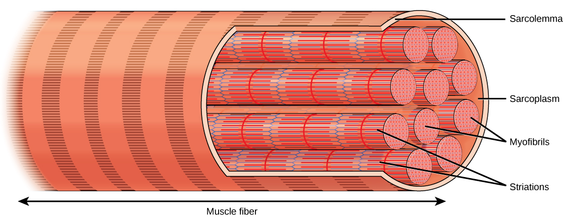
In skeletal muscle, thousands of sarcomeres, arranged end to end, form long tubes called myofibrils. These tubes are wrapped together in connective tissue and make up the bulk of the skeletal muscle cell (muscle fiber). Due to the length of each muscle fiber, many nuclei are scattered along the length of the cell. The average skeletal muscle cell is about 3 cm in length, but they can vary from about 1 mm (stapedius muscle of the middle ear) to over 50 cm (sartorius muscle of the leg).
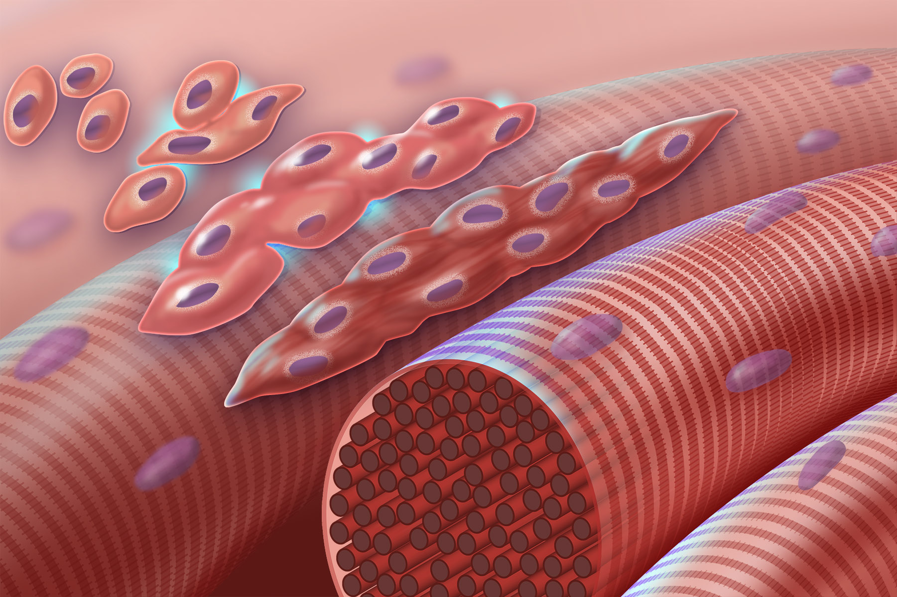
Cardiac muscle has a similar appearance to skeletal muscle because it is comprised of sarcomeres, however each cardiac muscle cell displays a branching appearance instead of the long, narrow tube-like appearance of skeletal muscle cells. The purpose of cardiac muscle contraction is to squeeze the heart and push blood out of the chambers and into the arteries, capillaries, and veins of the body with just enough force to return it back to the heart (Unit 16). A branching arrangement of cells allows for this type of movement. Like skeletal muscle, the coordinated shortening of thousands of sarcomeres results in the contraction of the heart muscle.
Cardiac muscle is unique in that it responds to signals that arise from specialized cells within the heart itself. These cells send electrical signals about every 0.8 seconds, causing the heart to contract about 72 times per minute (called heart rate or pulse). Under stress, hormones, nervous system signals, or both can cause the heart rate to increase. Similarly, signals from the body can slow the heart rate during periods of rest. The constant rate of contraction as well as the feedback loops involved in increasing or decreasing the heart rate are involuntarily controlled — lucky for us. Can you imagine having to consciously tell your heart to beat 72 times a minute? The penalty for forgetfulness would be severe.
With skeletal muscle, it is possible to contract a few individual fibers or entire groups of muscles all at once. With cardiac muscle, it is critical that all of the muscle fibers contract in unison. In order to achieve this, the heart muscle cells have a series of gap junctions that transmit the electrical signal for contraction all the way through the entire muscle simultaneously.
Structurally, the cardiac muscle needs to be strong so that it doesn’t rip itself apart with the constant, regular contractions. In order to do this, the heart has intercalated discs seen as zigzag lines on the surface of the drawing of cardiac cells above. These intercalated discs are regions with multiple desmosomes holding the muscle cells together. Remember that desmosomes are “spot-welds” that give a tissue structural integrity and strength.
Smooth muscle gets its name because it lacks the sarcomeres that give skeletal and cardiac muscle their striated appearance. Smooth muscle still makes use of actin and myosin proteins, but the arrangement appears more unorganized than in the other muscle types. The seemingly random arrangement of contractile proteins results in more of a squeeze-type contraction. This type of is more appropriate for moving substances throughout the digestive system or reducing the diameter of a blood vessel or the pupils of the eye. Smooth muscle contractions are typically slower to initiate, but last longer than other muscle types.
Smooth muscle is located in the walls of hollow internal structures, such as blood vessels, airways, lining the digestive system and in most organs of the abdominal cavity. It is also seen in other areas like the skin, eyes and reproductive organs.
Smooth muscle is not under voluntary control. Blood vessels expand and contract, airways open and constrict, our pupils dilate and constrict, and our gut keeps things moving, even while we sleep or while we’re otherwise distracted with other things.
The nerves that innervate smooth muscle are there to regulate smooth muscle contraction rather than control it. That is, the nerve cells release chemicals that increase or decrease the overall activity level of smooth muscle, but they don’t control individual muscle cells as much as in skeletal muscle.
The digestive system has its own nervous system that helps coordinate and control contraction of large blocks of smooth muscle, to keep things moving from mouth to anus without needing the brain to tell it what to do. The brain is capable of making helpful suggestions, but it doesn’t run the show in smooth muscle.
Objective 10.3
10.3.1 Describe the levels of skeletal muscle organization, from individual cells to muscles to muscle groups.
10.3.2 Identify the connective tissue layers around cells, fascicles and muscles and describe the way in which a muscle attaches to a bone or other muscle.
10.3.3 Explain the role of T tubules and sarcoplasmic reticulum within the cell.
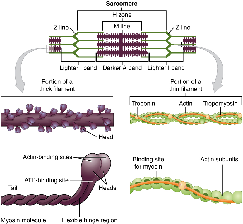
The smallest unit of skeletal muscle is the sarcomere, a regular arrangement of actin and myosin filaments plus structural proteins to hold things in place.
Many thousands of sarcomeres are lined up in a myofibril, a long strand of sarcomeres arranged end-to-end.
Several dozen myofibrils are found inside of each muscle cell, or muscle fiber. This is called a syncytium and is the result of embryonic muscle cells fusing to form a long tube.
The cell membrane of the skeletal muscle cell has the rather grand and strange name sarcolemma. It’s just like the cell membrane of any other cell; no one knows why this strange name persists.
Two Latin names appear over and over again in this Unit.
Sark or sarx (Greek σάρξ) means “flesh” in Greek, and so all the “sarco–” words come from that root: sarcoplasm, sarcolemma, sarcomere.
μῦς orMus, which became myo–, is the Greek word for mouse! The action of muscles like the biceps was thought to look like a mouse, probably by the same Greeks who named the constellations. They had a very active imagination.
Similarly, instead of the cytoplasm of muscle cells, we refer to the sarcoplasm.
muscle fiber = muscle cell
sarcolemma = cell membrane = plasmalemma
sarcoplasm = cytoplasm
sarcoplasmic reticulum = smooth endoplasmic reticulum
Lower motor neurons run throughout the muscle, but do not extend deep into each individual muscle cell. An elaborate system of tubules functions to relay the signal from the neuron resting just above the cell membrane to all of the sarcomeres inside the cell. These tubules, called T tubules, are like little folds of plasma membrane that dip into the cell. Flanking the T tubules on each side are tiny bags of calcium known as sarcoplasmic reticula. The sarcoplasmic reticulum is a specialized form of smooth endoplasmic reticulum which stores calcium ions (Ca2+) and releases them when needed. They are necessary in the contraction process.
Because the electrical and calcium management systems of the muscle cell must work together, as we’ll see later, they are in close proximity to each other. One cylindrical T tubule is flanked by two flattened sacs of smooth endoplasmic reticulum in a standardized arrangement called a triad.
In order to function properly, the contractile machinery inside muscle cells must be connected to tendon and bone. This connection is made by several levels of connective tissue. Each of these connective tissue layers ends in –mysium, which means “muscle”.
- Endomysium is a thin sheet of connective tissue that surrounds each muscle cell, lying just over the muscle cell, lying just over the muscle cell membrane (sarcolemma). This innermost connective tissue sheet has the prefix endo– which means “inner”.
- Perimysium is a connective tissue sheet which wraps about a dozen individual muscle cells. These groups of muscle cells, covered by perimysium, are called fascicles and are the smallest visible unit of muscle, seen as the “grain” in fresh meat. The prefix “peri–” means “surrounding”.
- Epimysium is a strong, thick sheet of connective tissue that covers an entire muscle. This is the sheet of dense irregular connective tissue that transitions into the dense regular connective tissue of the tendon. The prefix “epi–” means “on top of”, as in, it is a layer that is on top of the entire muscle.
Sometimes the connective tissues of muscle form broad, flat sheets that allow for muscles to attach across wide bone surfaces or to other muscles. These structures are called aponeuroses. Aponeuroses in the body can be found throughout the body, for example; where muscles attach to the skull, where the diaphragm attaches to the central tendon, and anywhere where larger muscles like the abdominal or trapezius muscles make connections. Tendons and aponeuroses have few blood vessels and nerves and therefore appear glossy and whitish in color.
Objective 10.4
10.4.1 Describe the organization of a sarcomere.
10.4.2 Diagram the location of myosin and actin filaments.
10.4.3 Explain what is meant by “sliding filament model.”
We have already discussed the way in which thousands of sarcomeres shorten to allow myofibrils to shorten, which collectively shortens the muscle cells, which shortens fascicles, which shorten muscles, giving us contraction. But how do the sarcomeres actually shorten? This objective will explain the way in which individual sarcomeres are able to shorten and return to their original shape.
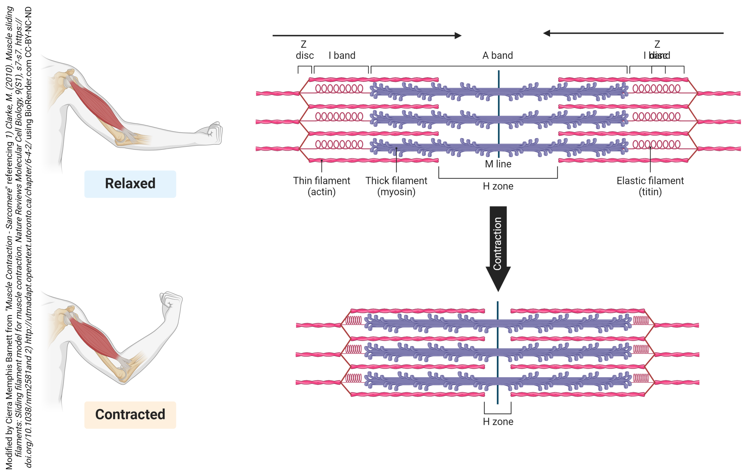
We saw in light micrographs of skeletal muscle that the striations that give striated muscle its name were sarcomeres, the contractile unit of muscle. The alternating areas of light and dark come about because of the arrangement of actin and myosin within each sarcomere.
The actin filaments are held together at the Z disc (line) by protein called actinin. The Z-disc is the dark line that splits the light areas in the micrographs seen here. The Z disc defines the borders of the sarcomere. Using electron microscopy, we can see the relationship between the thick filaments of myosin and the thin filaments of actin. There are regions where only actin filaments are found near the Z-discs, regions where only myosin filaments are found near the middle of the sarcomere or the M-line, and regions where they overlap. These regions changes as the sarcomeres shorten or spring back to their original shape after contraction.
We will see in more detail later how the centrally located myosin proteins reach across the gap and bind to the actin proteins and pull them towards the center of the sarcomere. As more myosin heads bind and pull in on actin, the sarcomere continues to decrease in length. After contraction, the actin binding sites on actin are covered and structural proteins like titin and elastin help return the sarcomere to its original shape. The process is repeated every time a muscle contracts. This is referred to as the sliding filament model.
It is important to note the extent of overlap between the actin filaments and the myosin filaments. The amount of overlap has a direct impact on the strength, or tension that can be generated by the contracting sarcomere. This relationship is known as the length-tension relationship.
Let’s start in the middle of the graph, where the most force can be generated. Notice the green lines representing the actin thin filaments, and the brown hairy thing representing the myosin thick filaments. The “hairs” are the myosin heads. In the middle, at a sarcomere length of about 2.5 μm, there are lots of opportunities for myosin to interact with actin (lots of potential points of contact between the brown “hairs” and the green lines).
Now look to the right side of the graph. If the sarcomere is stretched out to about 4 μm, then there is no overlap between the thin and thick filaments and no opportunity for myosin to generate force.
The hardest (and probably least important) to grasp is the left side of the graph. Here, the actin thin filaments overlap with each other; this also reduces the ability of myosin and actin to interact and generate force.
Muscle force, as we will see in Objective 6, is dependent on the interaction between actin and myosin molecules.
Objective 10.6
10.6.1 List the sequence of events from an increase in calcium in the muscle cell to contraction of the muscle.
The action potential in a muscle initiates contraction through a series of chemical reactions and protein interactions. Pay attention to the names of the key players in this objective and the role they play in shortening the sarcomere.
The calcium released from the sarcoplasmic reticulum interacts with a special protein, found only in muscle cells, called troponin. When calcium binds to troponin, it changes its shape, and pushes another specialized protein, tropomyosin. Tropomyosin normally covers the myosin binding sites on the actin molecule, preventing myosin heads from attaching and pulling in on the actin.
When calcium is present, troponin shoves tropomyosin, moving it away from the binding sites, which allows myosin to bind to the actin. This binding of myosin to actin starts a process called the cross-bridge cycle. It is called this because a link between myosin and actin forms, then breaks, then forms again, over and over.
It’s a cross-bridge cycle, so we can start anywhere, but let’s start at the top of these diagrams, at 12 o’clock on an imaginary clock face.
We start as the previous cross-bridge cycle has just completed, with the myosin head bound to the actin filament.
ATP is required for this process. When ATP binds to the myosin head, it releases the actin filament. People who are recently dead have no available ATP in their muscles. Because of this, we remain stuck at 12 o’clock in this diagram, with myosin bound irreversibly to actin. This biochemical condition is called rigor mortis when we see it in an entire human body.
At 3 o’clock, ATP binds to the myosin head and it releases the actin filament.
At 5 o’clock, ATP is split. The energy is stored in the myosin head for later use. Right now, the splitting of adenosine triphosphate (ATP) into adenosine diphosphate (ADP) and inorganic phosphate (Pi) results in a conformational change in the myosin head, converting it from a “flexed” (bent) position to an “extended” (straight) position.
At 8 o’clock, calcium comes in and causes a conformational change in troponin, which shoves the tropomyosin, exposing the myosin-binding site on actin molecules in the thin filament. Myosin, still in its “extended” position, grabs onto the actin thin filament.
At 10 o’clock, ADP and Pi leave the myosin head. Now we use the previously-stored energy from ATP. In the power stroke, the myosin head (still bound to actin) changes shape as ADP and Pi leave. As myosin heads “flex” (bend), the thick filament myosin heads slide the thin filament actin molecules along, generating force as the sarcomere shortens.
Notice the actin filament moving to the left in this diagram as the purple thick filament stays in one place. This is the connection to the sliding filament model seen earlier (Objective 4). 10-26
If the signal from the lower motor neuron stops, the calcium is actively pumped back into the sarcoplasmic reticulum. When this happens, troponin and tropomyosin return to their original location blocking actin binding sites. Without anything to bind to, myosin pulls away from the actin and the proteins mentioned earlier allow for the sarcomere to return to its normal, resting length.