Production, Circulation, and Resorption of Cerebrospinal Fluid
Jim Hutchins
Objective 2: Describe the formation of cerebrospinal fluid (CSF) and explain how it is stored and circulated in the CNS.
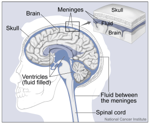
The brain has a series of hollow spaces. These are called ventricles.
Together, the ventricles and subarachnoid space are filled with cerebrospinal fluid (CSF), a filtrate of blood plasma that cushions and supports the brain, as well as supplying a favorable ionic environment for neurons (that is, isotonic and mostly NaCl). Scientists estimate that the brain, at an average 1400 g, weighs only 50 g when buoyed by CSF.
Cerebrospinal fluid is made in the choroid plexus, a specialized tissue that is found mainly in the lateral ventricles and fourth ventricle. There, the blood is filtered by ependymal cells, a type of glial cell.
CSF is almost identical to plasma; it is slightly lower in glucose and amino acids than plasma, and much lower in protein.
CSF circulates and is turned over about four times a day. It can be sampled with relatively little risk to the patient from the lumbar cistern, a large sac containing the cauda equina floating in CSF which is accessed from the intervertebral space between L2 and L3. Because of the circulation of CSF, a lumbar puncture may detect bleeding that originated (for example) in the subarachnoid space around frontal cortex.
The resorption of CSF is thought to occur in the arachnoid villi, a collection of structures along the midline of the superior aspect of the skull.
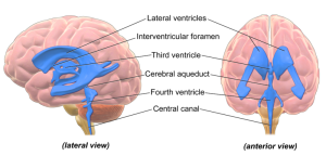
The ventricles include:
- Lateral ventricles, shaped like the letter C with a tail. This pair of ventricles is located beneath the white matter of cerebral cortex. There is no “first” or “second” ventricle, but there should be!
- A third ventricle, which is a slit-like space between the two eggs of the thalamus.
- A fourth ventricle, between the brainstem and cerebellum.
The ventricles are connected to each other by apertures or foramina. The lateral ventricles and third ventricle connect via the interventricular foramina. The third and fourth ventricles are joined by the cerebral aqueduct. The fourth ventricle communicates with the subarachnoid space surrounding the brain and spinal cord via three apertures: two lateral apertures and a single median aperture.
Hydrocephalus can also result from overproduction of CSF, or from slow resorption of CSF. Remember from an earlier objective that CSF can be sampled from the lumbar cistern, the subarachnoid space which contains the free-floating nerve roots of the cauda equina at the caudal end of the spinal cord.
Media Attributions
- Ventricles © Hoofring, Alan is licensed under a Public Domain license
- Ventricles Brain © BruceBlaus is licensed under a CC BY (Attribution) license
