Parasympathetic Nervous System
Jim Hutchins
Objective 3: Discuss the key features of the parasympathetic division of the autonomic nervous system.
General Features
Motor systems require an effector. The output of the nervous system is either contraction of a muscle or changing the secretion of a gland.
The somatic motor system contracts skeletal muscle. The autonomic motor system (visceral motor system) contracts smooth muscle, contracts cardiac muscle, or changes the secretion of glands.
The sympathetic and parasympathetic nervous systems have different anatomy and use different signaling molecules.
Both pathways consist of two neurons: a preganglionic neuron, with its cell body in the central nervous system and its axon terminals in an autonomic ganglion; and a postganglionic neuron, with its cell body in an autonomic ganglion and its axon terminals sending a chemical signal to the effector organ (cardiac muscle, smooth muscle, or a gland).
Parasympathetic Nervous System
| Parasympathetic Nervous System | |||||
|---|---|---|---|---|---|
| Preganglionic cell body location | Length of preganglionic axon | Preganglionic neurotransmitter (signaling molecule) | Postganglionic cell body location | Length of postganglionic axon | Neurotransmitter (signaling molecule) released onto effector organ |
| Brain stem or lateral horn of sacral spinal cord | Long | Acetylcholine | Ganglion near organ or in organ (intramural) | Short | Acetylcholine or nitric oxide (NO) |
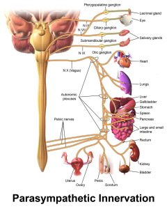 The anatomy, and therefore the physiology, of the parasympathetic nervous system is completely different. There is a long preganglionic neuron whose axon goes directly to the organ and a short postganglionic axon with the neuronal cell body generally found in the wall of the organ. The postganglionic neurons are found in clusters called intramural (“inside the wall [of the organ]”) ganglia. This allows the parasympathetic nervous system to control its targets organ-by-organ rather than body wide.
The preganglionic cell bodies for head, thoracic, and abdominal organs are found in the brainstem. Their axons travel in a cranial nerve (for the head) or in the vagus nerve (for the thorax and abdomen). Recall that the tenth cranial nerve is called the vagus nerve because it wanders all over the thoracic and abdominal cavities. As it wanders, it releases acetylcholine on the intramural ganglion neurons.
The preganglionic cell bodies for abdominal, retroperitoneal, and genital regions are found in the sacral spinal cord. They form pelvic splanchnic nerves which go to the digestive organs (abdominal), kidneys (retroperitoneal), bladder, and genitals.
The postganglionic neurons have cell bodies in the intramural ganglia. Their axons release acetylcholine, but this time, the same neurotransmitter acts on a different type of receptor, called the muscarinic acetylcholine receptor.
The parasympathetic nervous system is sometimes called the “rest and digest” system.
Compare and Contrast Sympathetic vs Parasympathetic Nervous Systems
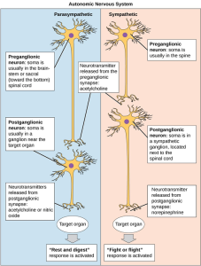
This diagram summarizes the features of the sympathetic and parasympathetic nervous system that we have discussed to this point.
Media Attributions
- Parasympathetic Innervation © BruceBlaus is licensed under a CC BY (Attribution) license
- Sympathetic vs parasympathetic © CNX OpenStax is licensed under a CC BY (Attribution) license
