Motor Cortex
Caleb Bevan
Objective 7: Identify the location of motor cortex, and the function of different motor cortical regions.
Motor Cortex
The areas of the cerebral cortex that are responsible for producing conscious, planned movement were introduced earlier, as we were subdividing cortical real estate based on the Brodmann classification. Areas responsible for movement, and their Brodmann numbers, are all in the frontal lobe. They include:
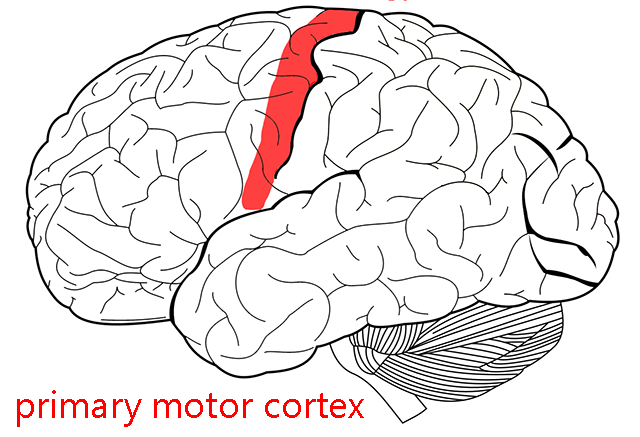
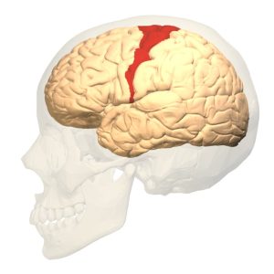
- Area 4: (precentral gyrus): the primary motor cortex, sending axons to the α motor neurons of spinal cord (executing movement).

-
- Area 6: premotor cortex and supplementary motor area (planning or imagining movement). Among the interesting neurons found here are mirror neurons, which are only active when watching someone else perform an action that you are trying to plan.
- Area 8: Frontal eye fields. This is the area of premotor cortex that plans eye movements.
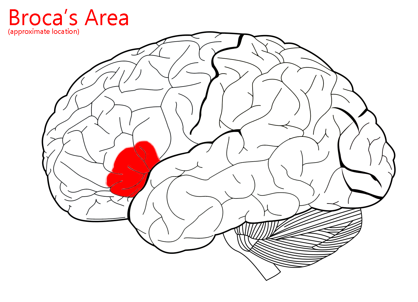
- Areas 44 and 45: Broca’s area. In most people, Broca’s area on the left side is responsible for the production of symbolic language. This can include speech, sign language, or writing. A lesion in this area produces aphasia.
For the time being, we will focus on the primary motor cortex (Brodmann 4 or precentral gyrus).
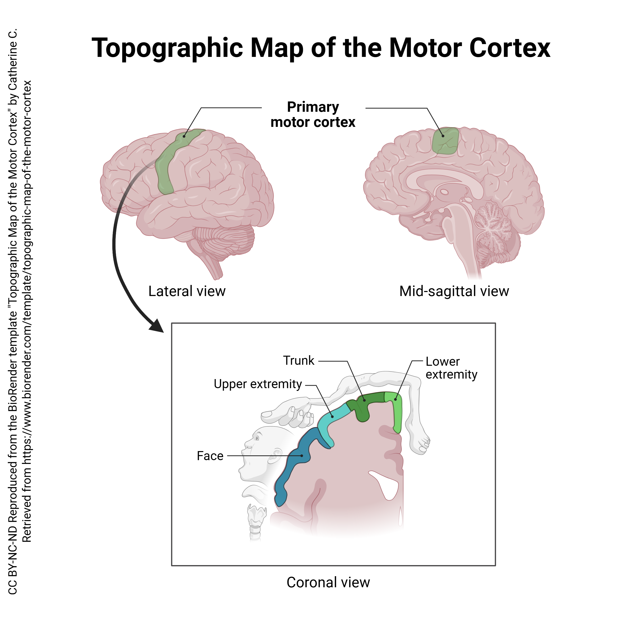
As for other motor and sensory areas of the brain, neurons which control movement form an orderly map of the body surface within the brain. This orderly map is called a homunculus (“little man”).
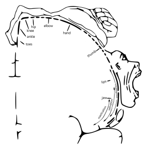 The motor homunculus, pictured here, is very similar to the sensory homunculus. The face areas are found in the most lateral part of the precentral gyrus. As we move towards the midline, we encounter, in order: hand, arm, shoulder, trunk, hip; in the medial longitudinal fissure, the map continues with leg and toes.
The lateral surface of the brain is supplied by a different set of arteries and arterial branches than the medial surface of the brain. Recall that the right side of the brain controls the left side of the body and vice versa. Therefore, a stroke (infarction, loss of blood supply) in the right lateral part of the precentral gyrus would produce paralysis of the left side of the face while a stroke in the right medial part of the precentral gyrus would produce paralysis of the left leg.
Information about conscious, willed movement is planned in the supplemental and premotor cortices. These areas then transfer their plan to the motor cortex, which convenes a neural “committee” to “vote” on which axons are going to be activated, and how many nerve impulses will be sent through each axon. The pyramid-shaped cells in layer V of area 4 (precentral gyrus) are called Betz cells. Betz cells are the largest neurons in the human body, and at 0.1 mm across, the cell bodies are visible to the naked eye in stained tissue. Accordingly, these cells have axons that are among the largest (and fastest) in the human body.
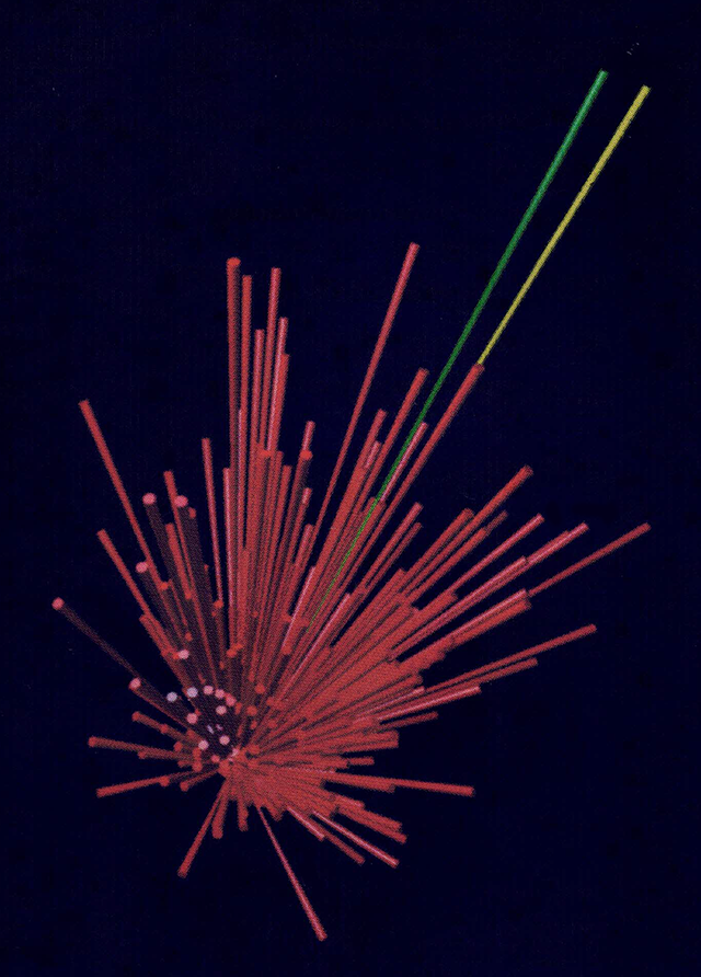
Apostolos Georgopoulos and colleagues have demonstrated how this vote occurs in an elegant series of experiments that beautifully illustrate the process. Rhesus monkeys were trained to push a red button when it lights up. The buttons were set on a panel in a row, an equal distance apart. To push them requires a movement at a known angle. Hundreds of cells in motor cortex fire at the same time, and because of the way they are wired to the output pyramidal neuron, the output pyramidal neuron generates a train of action potentials which activates the appropriate pool of motor neurons, each by the appropriate amount, in the anterior (ventral) horn of the cervical spinal cord gray matter. The activity of the output pyramidal neuron, and the direction of movement it causes, is shown by the green vector in the photograph above.
 The activation of the chosen pyramidal cell in motor cortex causes a contraction of the muscles of the arm and forearm that will approximate movement toward the desired target, in this case a lighted button. The movement that results is shown by a yellow vector. As described elsewhere, the slight mismatch between the preferred direction and force of contraction, and the actual direction and force of contraction, is corrected by the cerebellocortical circuitry.
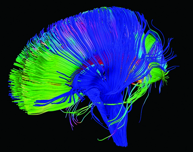
This image shows the motor axons, along with all other axons going to and from the cortex, in a structure that is collectively called the corona radiata (“radiating crown”). The technique shown here, diffusion tensor imaging, is useful in seeing when the Betz cell axons or any other axon bundles are damaged, as in concussion.
Premotor Cortex
The earliest research about the premotor cortex was done in 1905 by Dr. Campbell. He was the first one to suggest that the motor cortex might be separated into at least two different regions because of the analysis of the neural tissue itself (the premotor cortex is one of the many subdivisions of the motor cortex). After some time there was many disagreements and the loss of the term premotor cortex till the 1980s with many researchers providing more data in support of the idea of the premotor cortex. Overtime the other areas of the motor cortex were also found and studied to what we have now which is a plethora of areas in the motor cortex in charge of different functions/movements.
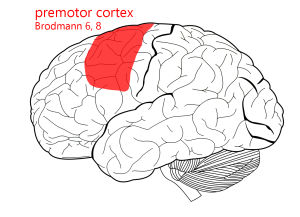
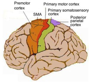
The premotor cortex is located posterior to the frontal lobe. The premotor cortex might also be referred too as Brodmann’s area 6 or 8 (see image above). Is is one of the main subdivisions of the motor cortex which is the main hub for motor signals from your brain.
There are two subdivisions of the premotor cortex; the dorsal premotor cortex and the ventral premotor cortex. Both are named by the sub location in the premotor cortex.
The dorsal premotor cortex (PMd) in primarily in charge of guiding reaching, complex movements of arm, use of sensory stimuli in movements, and relating to eye movement.
The ventral premotor cortex (PMv) is also in charge of sensory guidance of movement along with shaping hand/grasping of hand.
Both parts of the premotor cortex are constantly working together in almost all movement in the body most importantly the arm and hand.
-
-
-
- Cortical inputs: Prefrontal cortex, parietal lobe
-
Outputs: M1, brainstem, spinal cord
-
Outgoing connections: motor cortex, spinal cord, basal ganglia, cerebellum
-
-
Many functions of the premotor cortex are still not fully understood to this date. There is research to support that the premotor cortex is used for things including spatial guidance in movement, planning out movement, and observing the actions of others. They use all of these things to make a movement that is appropriate for the situation.
Motor Planning and Preparation
-
-
-
- Role in preparing movements before execution
-
Sequencing of motor acts
-
-
Sensorimotor Integration
-
-
-
-
Processing of visual and sensory inputs to guide movement
-
-
-
Goal-Directed Movements
-
-
-
-
Reaching, grasping, and tool use
-
Movement selection based on cues
-
-
-
Motor Learning and Skill Acquisition
-
-
-
-
Role in associative and observational learning
- Watching others and putting into practice the movement
-
-
-
Complex movements such as reaching or climbing demand greater coordination, the integration of more control variables, the tracking of objects in the nearby environment, and the ability to plan actions seconds ahead. In contrast, simpler components of the motor system—like moving an object with your fingers after it has been grasped, or using your mouth to manipulate objects—require less foresight, reduced spatial computation, and rely more on fine control of joint movements and muscle fibers.
According to this, more complex, especially multi-joint, movements are represented more prominently in the anterior regions of the motor cortex. This area of the cortex tends to prioritize control over the back and neck, which functions as a key figure in bodily coordination. Simpler motor acts that primarily involve further muscles are emphasized further posteriorly within the motor cortex.
Interpretation suggests that while primary motor cortex is more involved in relatively simple movements and the premotor cortex in more complex ones, this spatial distinction does not indicate a control hierarchy. Rather, these regions are functionally distinct and internally diverse, reflecting the natural movement range of our bodies.
Supplementary Motor Area
The supplementary motor area or SMA is the dorsal medial part of the frontal lobe. Neurons in the SMA apply directly to the neurons in your cortical spinal tract. There has been research to show they have a role in voluntary movement, stabilization of the body, and bimanual coordination. There is no affect from stimulation in the SMA and all movement is internally generated. Graziano and colleagues have looked at SMA and emphasis in locomotion (the ability for an organism to move from one place to another). They looked at the use in things like climbing or jumping and created a hypothesis that the SMA is related to locomotion.
Functional Roles
Pre-SMA
-
-
- Most anterior portion. It has no connections to spinal cord or motor cortex. Has connection in prefrontal areas.
-
Inputs: Basal ganglia, thalamus, prefrontal cortex
-
Outputs: M1, spinal cord (via corticospinal tract)
-
- Most anterior portion. It has no connections to spinal cord or motor cortex. Has connection in prefrontal areas.
-
SMA proper
-
-
- The SMA proper is located at the top of the hemisphere partly on the medial well. Has been thoroughly researched in monkeys. One of the primary outputs of the cortical motor system through the spine.
- Inputs: Same as Pre-SMA
-
Outputs: Same as Pre-SMA
- The SMA proper is located at the top of the hemisphere partly on the medial well. Has been thoroughly researched in monkeys. One of the primary outputs of the cortical motor system through the spine.
-
Supplementary Eye Field
-
-
- When stimulated causes head and eye movements.
-
Dum and Strick
-
-
- Connections to spinal cord and cingulate sulcus. Has not been thoroughly studied and is theorized to deal with emotions
-
Frontal Eye Fields
The inferior most part of Brodmann 8 premotor cortex has a specialized function, controlling the eye muscles. This area is called the frontal eye fields. Small movements called saccades are used to move the eyes to find a target. The frontal eye fields must carry out a swift and precise calculation to bring both eyes to the same location in visual space, so that binocular vision and depth perception can occur.
Media Attributions
- Primary motor cortex © Carter, Henry Vandyke adapted by Jim Hutchins is licensed under a CC BY-SA (Attribution ShareAlike) license
- Broca’s area © Carter, Henry Vandyke adapted by Jim Hutchins is licensed under a CC BY-SA (Attribution ShareAlike) license
- Topographic Map of the Motor Cortex © Catherine C is licensed under a CC BY-NC-ND (Attribution NonCommercial NoDerivatives) license
- Brodmann area 4 lateral © Anatomography is licensed under a CC BY-SA (Attribution ShareAlike) license
- Motor homunculus © Wilder Penfield is licensed under a Public Domain license
- Population coding motor cortex © Apostolos P. Georgopoulos is licensed under a All Rights Reserved license
- DTI corona radiata © P Basser NICHD is licensed under a CC BY (Attribution) license
- Premotor area 6 area 8 © Carter, Henry Vandyke adapted by Avalon Marker and Jim Hutchins is licensed under a CC BY-SA (Attribution ShareAlike) license
- Human motor cortex © Pancrat is licensed under a CC BY-SA (Attribution ShareAlike) license
