Lobes of the Human Brain
Jim Hutchins
Objective 1: Enumerate the lobes of the brain.
Important Locations Objective 1 Video Lecture
These subdivisions of the developing brain become adult structures, as shown in the table. Because each of these areas has a different anatomy and different strategy of “brain wiring”, it’s important to understand just a little bit about the development of the brain to understand the anatomy and function of the brain.
For example, the cerebral cortex and basal nuclei, which represent the vast majority of the weight of the human brain, are extensively interconnected and control much of human behavior. The basal nuclei smooth out the activity of the part of cerebral cortex that controls muscles; this smoothing goes wrong in Parkinson disease. Changes in the activity of another part of the basal nuclei is implicated in obsessive-compulsive disorder.
Basal Nuclei or Basal Ganglia?
Many authorities call the basal nuclei the basal ganglia. I believe this is confusing; a ganglion is a collection of nerve cell bodies in the PNS. The basal nuclei fit squarely within the CNS. That’s a good reason to go through the hassle of changing the name for our sons and daughters who might have to learn neuroanatomy later.
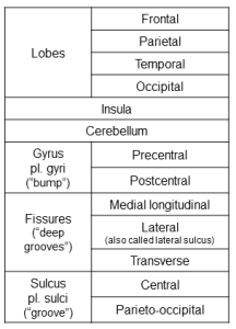
This table contains the names of all the brain structures we need to learn at this point in the course.
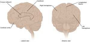
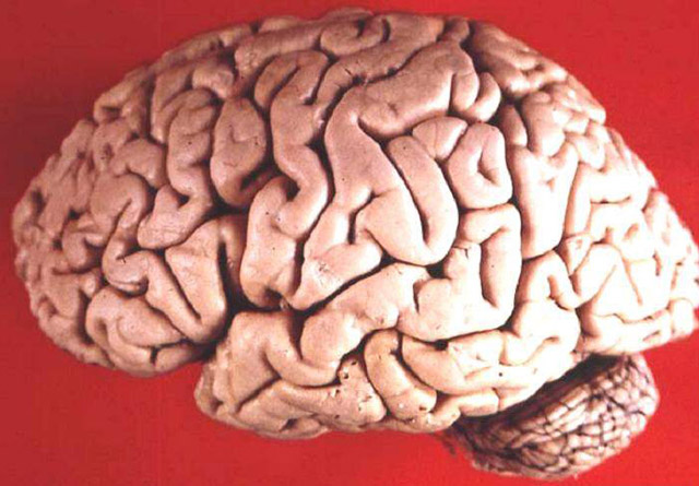
A deep groove, the medial longitudinal fissure, divides the brain into a left hemisphere and right hemisphere. These are connected by the corpus callosum, a band of axons running between mirror-image areas of the brain, right and left.
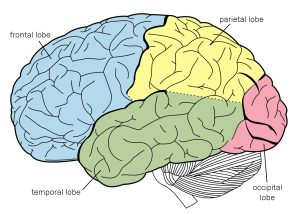
First, we’ll divide the external surface of the cerebral cortex into four lobes.
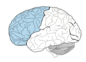
The frontal lobe (wait for it…) is in front. (left)
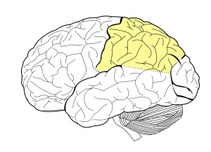
The parietal lobe shares its borders with the other three external lobes. It is just posterior to the frontal lobe. (right)
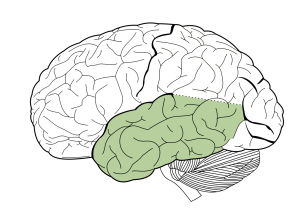
The temporal lobe (wait for it…) is under the temple. (left)
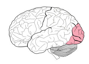
The occipital lobe is the most posterior lobe of the brain. (right)
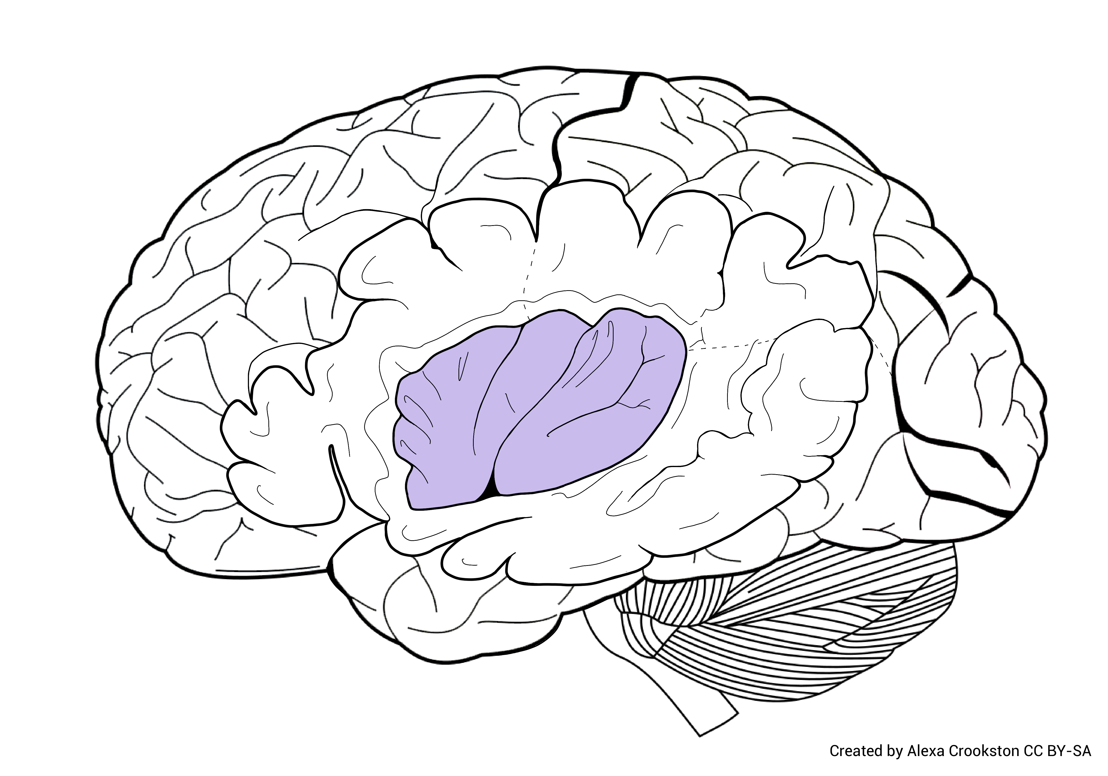 The insula is a fifth lobe (insular lobe). This is the part of the brain that is folded inside, and only visible when we cut the lateral surface of the brain away. Scientists believe the insula is the source of social emotions like guilt or pride. It may also be the area of the brain that interprets or drives cravings.
The cerebellum, attached to the brainstem, is a separate cortex (cerebellar cortex) with a separate structure.
Its job is to compare the motor programs of the brain to what the body is actually doing in real time and correct any mistakes that are being made. It is connected to the brainstem by three cerebellar peduncles (“little feet”).
Media Attributions
- Table of structures © Jim Hutchins is licensed under a CC BY-SA (Attribution ShareAlike) license
- Cerebrum © Betts, J. Gordon; Young, Kelly A.; Wise, James A.; Johnson, Eddie; Poe, Brandon; Kruse, Dean H. Korol, Oksana; Johnson, Jody E.; Womble, Mark & DeSaix, Peter is licensed under a CC BY (Attribution) license
- Lobes of the brain © Carter, Henry Vandyke adapted by Jim Hutchins is licensed under a Public Domain license
- Frontal Lobe © Carter, Henry Vandyke adapted by Jim Hutchins is licensed under a Public Domain license
- Parietal Lobe © Carter, Henry Vandyke adapted by Jim Hutchins is licensed under a Public Domain license
- Temporal Lobe © Carter, Henry Vandyke adapted by Jim Hutchins is licensed under a Public Domain license
- Occipital Lobe © Carter, Henry Vandyke adapted by Jim Hutchins is licensed under a Public Domain license
- Insula colored area © Carter, Henry Vandyke adapted by Alexa Crookston is licensed under a CC BY-SA (Attribution ShareAlike) license
- Human brain lateral view © John A Beal, PhD is licensed under a CC BY (Attribution) license
