Lacrimation
Jim Hutchins
Objective 7: Explain the process for making tears (lacrimation).
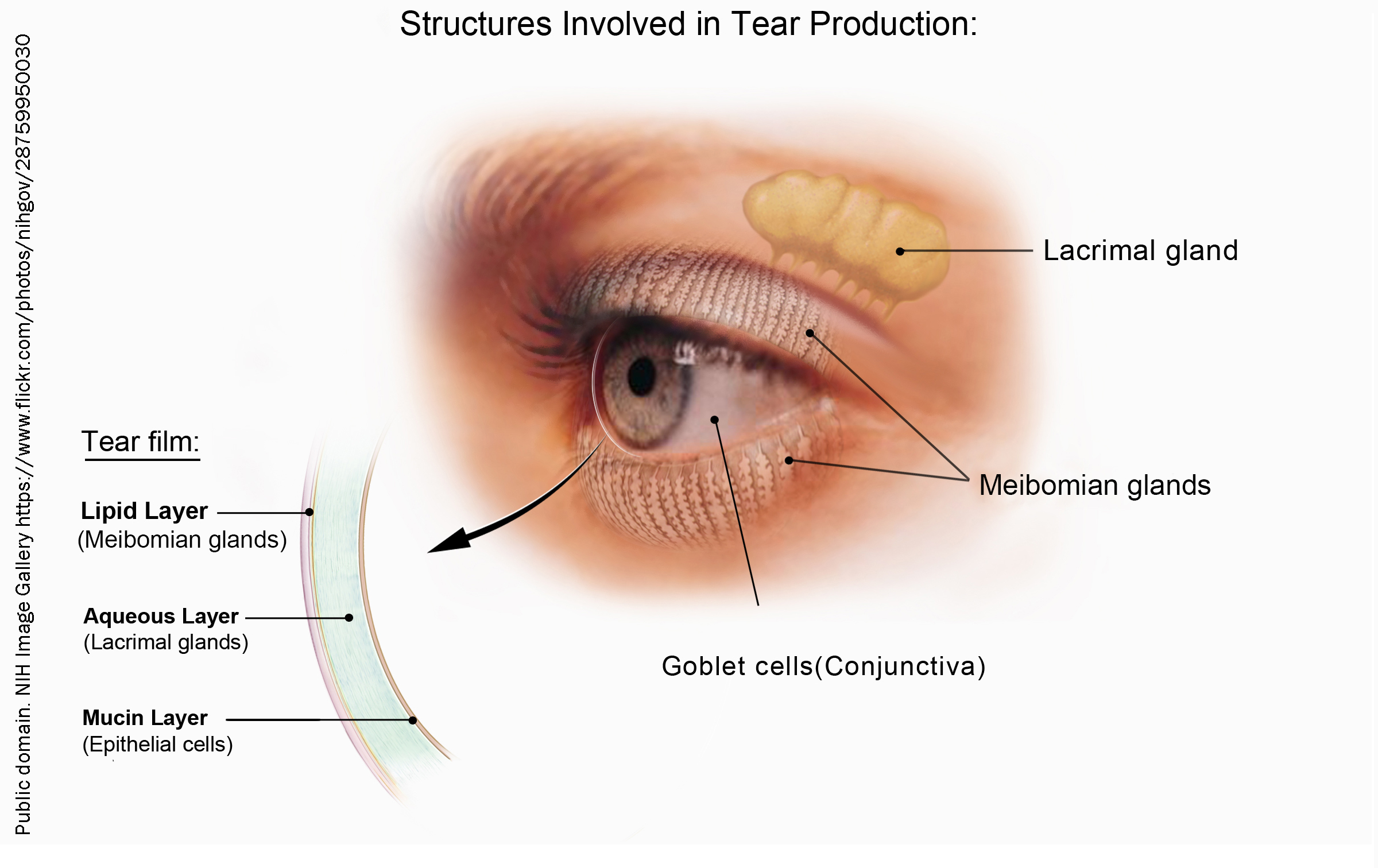
When we cry, or the eye is irritated, lacrimal glands in the orbit just above the superior eyelid are activated, along with Meibomian glands in the superior and inferior eyelid. Goblet cells found in the epithelium of the conjunctiva add mucus. Thus the tear film consists of three layers, from the conjunctiva out:
- a mucin layer made by the goblet cells;
- an aqueous (watery) layer contributed by the lacrimal glands; and
- a lipid layer contributed by the Meibomian glands.
The lacrimal glands add over 1500 proteins to the watery layer of the tear film, but the most prominent ones are:
- immunoglobulin A (for immune defense)
- lactoferrin
- protein G
- lipocalins
- prealbumin
- apolipoprotein D
- lysozyme
Lipocalins are a family of proteins that form an interface between the watery and lipid layers and help with surface lipid spreading. They may also help to remove harmful lipophilic molecules.
About 1.2 µL/min of fluid is continually produced by the lacrimal glands, but the rate of tear production can be massively increased by noxious stimuli or by psychological stimuli.
Emotional crying is unique to humans. Evolutionary psychologists believe it developed as a modification of the distress vocalization response (that is, a sort of silent scream). Neurons of the anterior cingulate cortex, ventromedial prefrontal cortex, basal nuclei, thalamus, hypothalamus, and amygdala all seem to be able to stimulate this response.
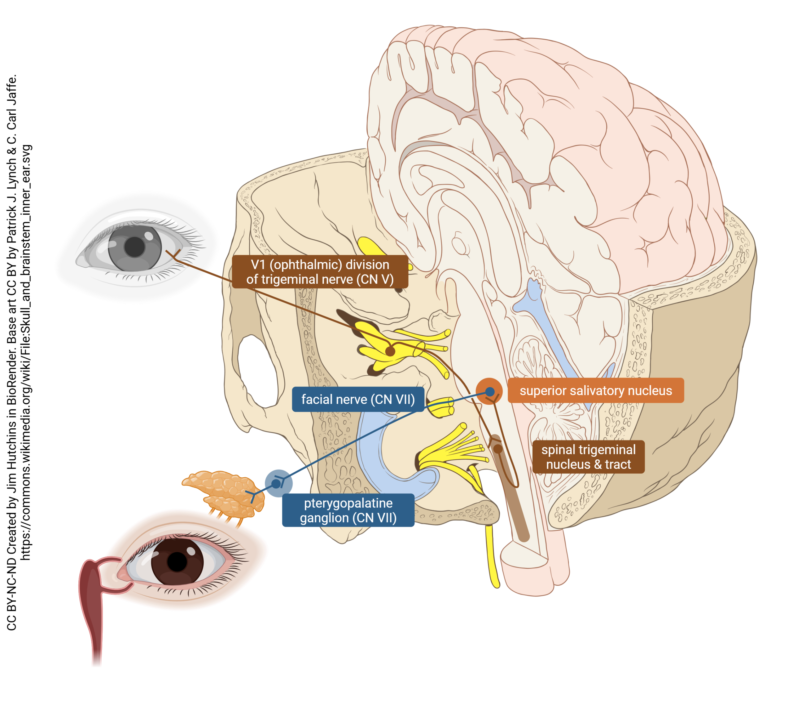
For lacrimation triggered by noxious stimuli delivered to the eye, the pathway is as follows. Sensory receptors (free nerve endings) are found in the conjunctiva. The sensory information is carried on the ophthalmic division of the trigeminal nerve (cranial nerve V); this branch of the trigeminal is usually designated V1. The cell body of these unipolar sensory neurons is in the trigeminal ganglion in Meckel’s cave, a depression in the temporal and sphenoid bones near the pons. These fibers enter the pons as the trigeminal nerve, and then descend to make a synapse in the spinal trigeminal nucleus, named this because it extends from the mid-pons down to the level of the foramen magnum, where the spinal cord begins.
The neuron carrying the sensory information sends an axon up the spinal trigeminal tract (which parallels the hot-dog-shaped nucleus) to synapse onto the preganglionic parasympathetic motor neurons found in the superior salivatory nucleus, the parasympathetic division of the facial nerve (CN VII) nucleus.
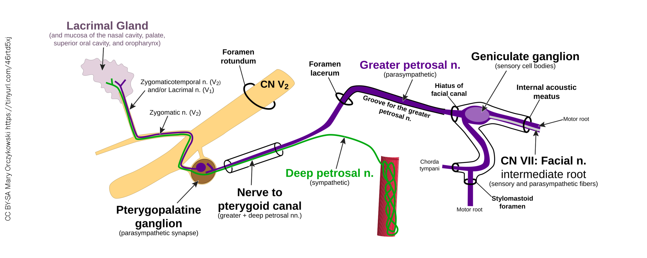
The preganglionic parasympathetic axons travel in the greater petrosal nerve to the pterygopalatine ganglion, where they synapse onto postganglionic parasympathetic neurons that send axons a short distance via the zygomatic nerve and/or lacrimal nerve to innervate the lacrimal gland and increase tear secretion.
As with other systems we think of as parasympathetic, there is a small contribution from the sympathetic nervous system. These long postganglionic axons travel along with blood vessels until they jump off forming the deep petrosal nerve which (anatomically but not functionally) joins the parasympathetic axons at the pterygopalatine ganglion.
Media Attributions
- Tear production © National Eye Institute, National Institutes of Health is licensed under a Public Domain license
- Lacrimal reflex pathway © Patrick J. Lynch and Carl Jaffe adapted by Jim Hutchins is licensed under a CC BY-NC-ND (Attribution NonCommercial NoDerivatives) license
- Autonomic Innervation to the Lacrimal Gland © Anatomary is licensed under a CC BY-SA (Attribution ShareAlike) license
