How the Neural Tube Develops Into the Major Divisions of the Central Nervous System
Jim Hutchins
Objective 2: Recognize the three primary and five secondary brain vesicles.
Nervous System Assembly Objective 2 Video Lecture
The vertebrate nervous system develops, just like every part of the body, from a single fertilized egg. In humans, about 16 days after the union of sperm and egg (conception), an event called gastrulation occurs. (Note that 16 days post-fertilization is the same as about 30 days from the last period, which is the method mosts doctors and patients use to measure human development.)
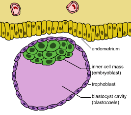
Before gastrulation, the vertebrate embryo is a hollow ball of rapidly dividing cells called a blastocyst, which means “a hollow ball of rapidly dividing cells” in Latin.
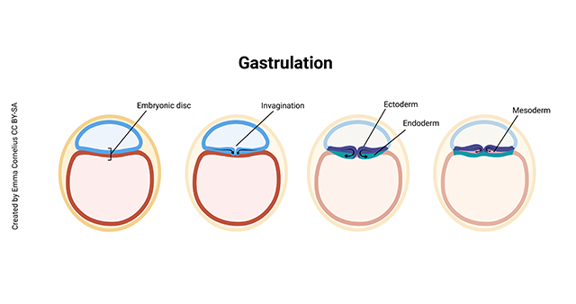
In the process of gastrulation, the hollow ball folds in on itself (think of poking your finger into an uninflated tennis ball). This forms three layers:
- ectoderm (Latin: “outer skin”)
- mesoderm (“middle skin”)
- endoderm (“inner skin”)
The ectoderm forms the skin and nervous system, which is why these two tissues are closely related to each other. For example, in the disease neurofibromatosis, there may be pigmented spots on the skin in the mild forms of the disease all the way to nervous system tumors in the more severe forms of the disease.
The ectoderm which is destined to be nervous tissue forms a structure called the neural plate.
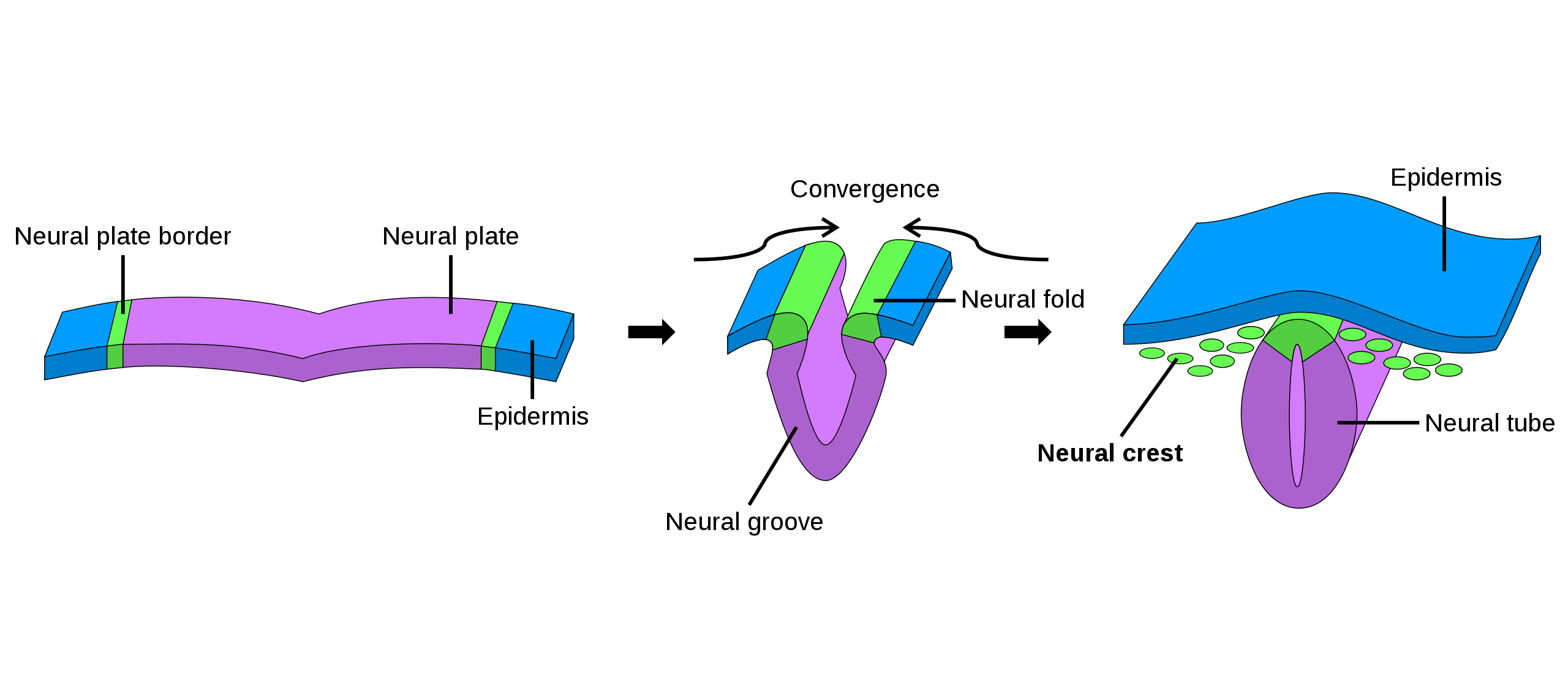
Under the influence of chemical signals from the underlying rod of tissue called a notochord, the neural plate cells take on a cone-like shape, which bends the entire plate into a groove which then gradually deepens. Finally, when the groove becomes as deep as it can possibly be, the dorsal walls meet and fuse to form a neural tube.

Once the neural tube forms, it starts to bend and pinch itself into three primary brain vesicles:
- prosencephalon (forebrain)
- mesencephalon (midbrain)
- rhombencephalon (hindbrain)
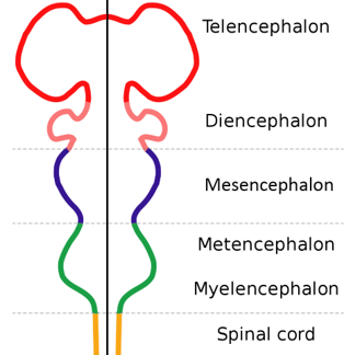
These three primary brain vesicles then continue changing shape to form five secondary brain vesicles:
- telencephalon
- diencephalon
- mesencephalon
- metencephalon
- myelencephalon
The prosencephalon gives rise to the telencephalon and diencephalon.
The mesencephalon (midbrain) stays the mesencephalon. This will be the smallest division of the adult brain.
The rhombencephalon gives rise to the metencephalon and myelencephalon.
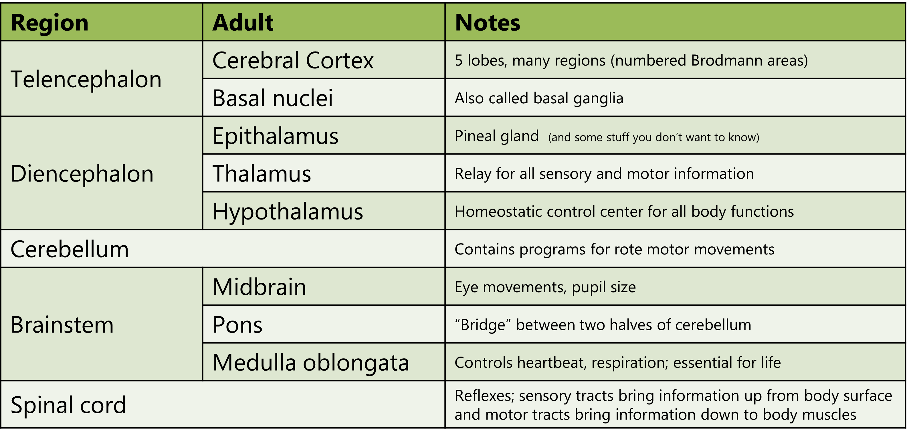
The table below shows the adult brain structures which develop from the five secondary brain vesicles (left column). In the “Notes” column, there is a bit more information about what each part of the brain does.
Media Attributions
- Blastocyst © Seans Potato Business is licensed under a CC BY-SA (Attribution ShareAlike) license
- Gastrulation © Emma Cornelius is licensed under a CC BY-SA (Attribution ShareAlike) license
- Neural crest © NikNaks is licensed under a CC BY (Attribution) license
- Brain Vesicles © Betts, J. Gordon; Young, Kelly A.; Wise, James A.; Johnson, Eddie; Poe, Brandon; Kruse, Dean H. Korol, Oksana; Johnson, Jody E.; Womble, Mark & DeSaix, Peter adapted by Jim Hutchins is licensed under a CC BY (Attribution) license
- Primary and secondary brain vesicles © Nrets adapted by Jim Hutchins is licensed under a CC BY-SA (Attribution ShareAlike) license
- Table of adult brain structures © Jim Hutchins is licensed under a CC BY-SA (Attribution ShareAlike) license

.svg)