Retinal Output: Ganglion Cells
Jim Hutchins
Objective 8: Demonstrate how retinal ganglion cells process information to produce a neural output signal from the retina.
The first thorough studies of retinal ganglion cell types were performed in the cat. The researchers discovered two types of response to sine wave gratings, which (according to legend) they called “interesting” and “uninteresting”. These initial classifications resolved into the more scientifically-defensible terms X (with linear spatial summation) and Y (with non-linear spatial summation). That is, X cells fire a pattern of action potentials that matches the sine wave grating, so there are densely-clustered action potentials near the peaks of the sine wave and sparse action potentials near the troughs of the sine wave. Y cells fire a burst when the wave rises, and another when it falls. The Y cells detect when the stimulus changes; thus, if you turn on a light, the Y cells respond with a burst and then go silent until you turn the light off, when they fire another brief burst. The physiological type “X” corresponds well to the morphological type “α” and the physiological type “Y” almost matches the morphological type “β”.
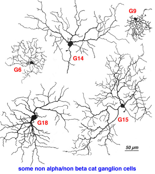
The above illustration shows drawings of ganglion cells that fit into neither category and are called non-α, non-β.
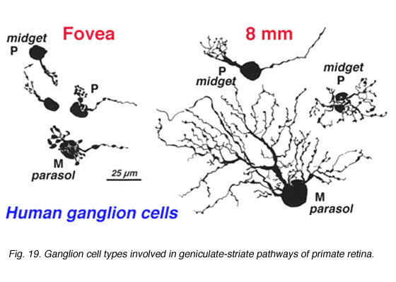
Human ganglion cells are classified into three groups. The naming is based on historical considerations and is a bit confusing, but here goes. The three types of ganglion cells are:
- M cells
- P cells
- non-M, non-P cells
In order to detect motion, retinal ganglion cells must collect information indirectly over a large portion of the retina. In particular, the ganglion cell is interested in what a large range of amacrine cells is reporting about change in change (i.e. moving edges). These ganglion cells have large dendritic trees supported by large cell bodies. They will eventually synapse onto large (magnocellular) cells in the LGN and are therefore called M ganglion cells.
In order to detect fine details, another set of retinal ganglion cells must restrict the areal extent of their inputs, collecting information indirectly from a small group of photoreceptors, horizontal cells, and amacrine cells, and directly from perhaps only one bipolar cell. This means the ganglion cell dendritic tree is small, and it is supported by a small cell body. Eventually, these will synapse onto small (parvocellular) cells in the LGN, and for this reason these ganglion cells are called P ganglion cells.
Ganglion cells which will contribute to color processing at higher levels are dumped in the bin labeled non-M, non-P.
As we will see, this theme of specialized cells that are most interested in motion and specialized cells that are most interested in details of form will continue throughout the visual system.
Ganglion cells will convey all this information to the LGN. Since they are the only cells that send information over long distances, they are the only cells that fire action potentials. All the other types of retinal cell use graded potentials, with small fluctuations in neurotransmitter release, to convey information. (Although some amacrine cells use calcium spikes, these are not technically action potentials.)
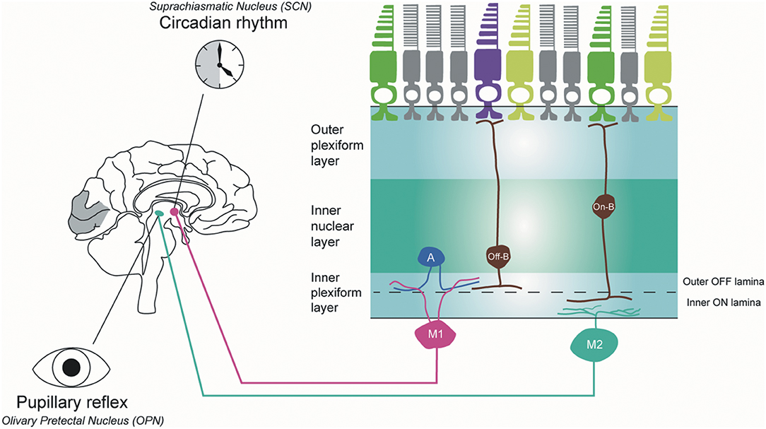
There are a special category of ganglion cells that have a huge dendritic tree covering a significant portion of the entire retina. These cells, the melanopsin intrinsically photosensitive ganglion cells, are responsible for telling the visual system when the sun rises in the morning and when it sets at night. That is, they respond best to changes in light not in a defined spot but over the entire visual field. These cells are important in maintaining circadian rhythms tied to day/night cycles and in the pupillary light reflex. Thus, these ganglion cells don’t send axons to the lateral geniculate but rather to the suprachiasmatic nucleus in the hypothalamus and to the pretectal olivary nucleus (near the oculomotor CN III nucleus) in the midbrain.
Color Processing in Retinal Ganglion Cells
We’ve looked at lateral interactions mediated by horizontal cells in the OPL and amacrine cells in the IPL. There is a straight-through pathway with less lateral interaction and this pathway tends to code for color. A cone photoreceptor will synapse extensively onto a so-called cone bipolar (also carrying the evocative name “midget bipolar”) which in turn passes information to the dendrites of a ganglion cell.
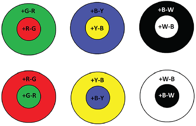
Ganglion cells which receive information via bipolar cells from cone photoreceptors have color-opponent receptive fields. That is, if the receptive field center depolarizes to red light, the receptive field surround hyperpolarizes to green light. If the receptive field center hyperpolarizes to yellow light, then the surround depolarizes to blue light.
Media Attributions
- Ganglion cells cat non-alpha non-beta © Helga Kolb, PhD. is licensed under a CC BY-NC (Attribution NonCommercial) license
- Ganglion cells human © Helga Kolb, PhD. is licensed under a CC BY-NC (Attribution NonCommercial) license
- Ganglion cell morphology © Ungsoo Samuel Kim, Omar A. Mahroo, John D. Mollon, and Patrick Yu-Wai-Man is licensed under a CC BY (Attribution) license
- Ganglion cell responses © Ungsoo Samuel Kim, Omar A. Mahroo, John D. Mollon, and Patrick Yu-Wai-Man is licensed under a CC BY (Attribution) license
- Melanopsin intrinsically photosensitive ganglion cells © Ungsoo Samuel Kim, Omar A. Mahroo, John D. Mollon, and Patrick Yu-Wai-Man is licensed under a CC BY (Attribution) license
- Color opponent receptive fields © Aristeidis Tsitiridis, Cristina Conde, Beatriz Gomez, and Enrique Caballo is licensed under a CC BY (Attribution) license
