Electrical Synapses
Caleb Bevan
Objective 5: Illustrate the properties of electrical synapses.
We have been focused on chemical synapses as a source of information transferred from one cell to another. Another type of synapse, which transfers information bidirectionally, is based off a structure found in many different cell types, not just neurons.
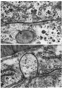
In non-neuronal cells, this type of connection between cells is called a gap junction. This name came from the narrowing of the space between cells seen in electron micrographs. In this electron micrograph, we are looking at two liver cells at the left and right of the electron micrograph. In the middle are the cell membranes, seen as dark lines. Note how the lines are separated at the top and bottom but between the barely visible black arrows, there is a gap junction.
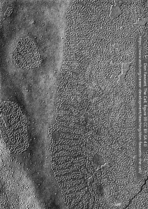
When electron microscopists use a special procedure called freeze-fracture, the gap junction appears as a series of bumps with holes in the center, like a sheet pan of donuts. Here, we are looking at a gap junction in a granulosa cell of the rat ovary.
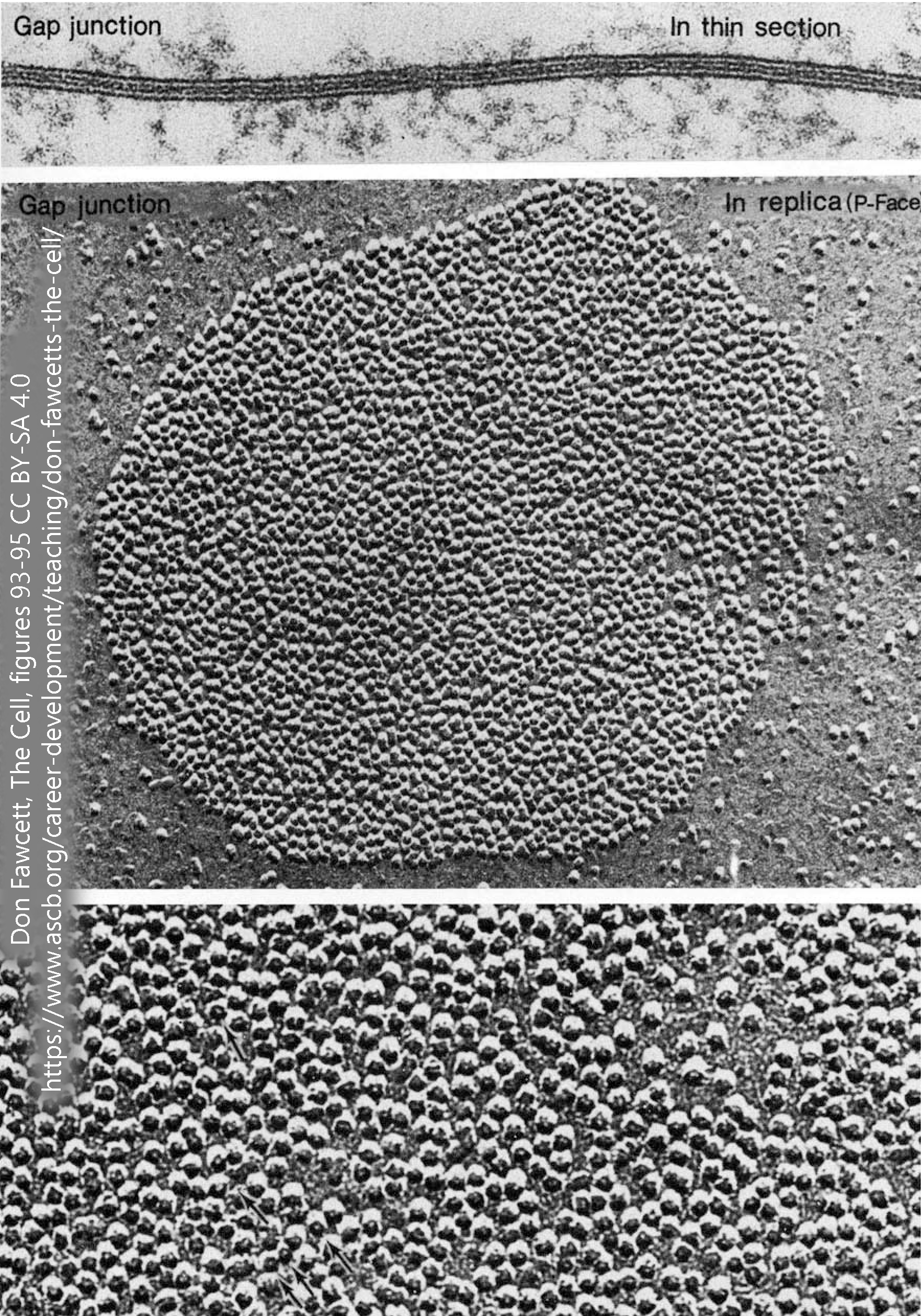 We have to blow these pictures up really big to see the donut hole, however. It appears as a white dot in the bottom freeze-fracture image. The whole channel (“donut”) is 7 nm across. The hole is about 2 nm (0.000002 mm, or about 20 hydrogen atoms), just big enough to let through molecules of about 1000 Da molecular weight. For comparison, Na+ is 23 Da; Ca2+ is 40 Da; glucose is 180 Da; and ATP is 507 Da; all of these will fit through a hole this size.
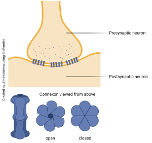 Electrical synapses are the neuronal equivalent of the gap junction and are structurally indistinguishable from gap junctions found in non-neuronal cells. The “donuts” we saw earlier are a protein called connexin; six connexin protein subunits from one cell, and six connexin protein subunits from a second cell, are all joined together to form an adaptable pore called a connexon.
Connexons are normally open. This allows ions such as Na+, K+, and Cl– to pass easily between the cells. We learned earlier that action potential propagation results from the spread of Na+ and K+ ions inside the cell, and so (for example) an action potential can pass with only a small amount of difficulty between two cells this way. In general, if an action potential is generated by the top cell in this diagram, then the voltage change in the bottom cell will be large enough to reach threshold and trigger another action potential in the bottom cell.
This setup allows cells to synchronize their activity, which is important in animals that need a strong escape response. For example, a cockroach will respond to a visual stimulus such as a rolled-up magazine with an escape behavior.
But the disadvantage of this approach is that the synapse cannot “learn” or modify its behavior. In the next section of the course, we’ll look at modifiable synapses and see how these produce a much more effective nervous system. For this reason, mammals such as humans have relatively few electrical synapses, although they do exist.
Another important feature of a connexon is its ability to seal off and thereby cut off the electrical synapse. In general, this happens because Ca2+ rises in one cell. Increased Ca2+ is a good indicator that the cell is dead or dying, and so it’s important not to share ions with sick or dead cells — substances which can pass through the connexon can kill you, too.
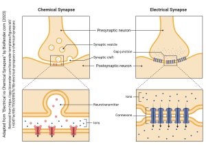 This figure compares and contrasts the chemical synapse and electrical synapse and also reminds us of the salient features of each.
Media Attributions
- Gap junction © Richard Wood is licensed under a CC BY-NC-ND (Attribution NonCommercial NoDerivatives) license
- Gap junctions © Don W. Fawcett is licensed under a CC BY-NC-ND (Attribution NonCommercial NoDerivatives) license
- Gap junctions saccular macula goldfish © Kiyoshi Hama and David Albertini is licensed under a CC BY-NC-ND (Attribution NonCommercial NoDerivatives) license
- Connexons and the electrical synapse © Jim Hutchins is licensed under a CC BY-NC-ND (Attribution NonCommercial NoDerivatives) license
- Comparison of electrical vs chemical synapses © BioRender adapted by Jim Hutchins is licensed under a CC BY-NC-ND (Attribution NonCommercial NoDerivatives) license

