Color Processing in the Retina
Jim Hutchins
Objective 3: Relate how the three cone types of the human retina contribute to color vision.
Photons are particle-waves. This means they sometimes act as particles (such as when we talk about refraction of light rays by the cornea and lens) and sometimes act as waves (such as when we talk about their wavelength, or when we do an experiment called the two-slit experiment). The realization that light has this dual quality only came about at the turn of the 20th century and was truly a paradigm shift in our understanding of vision.
The different wavelengths of light possess different energy. This energy, which has both electrical and magnetic qualities, is accordingly called electromagnetic energy. An entire spectrum of electromagnetic energy exists, from the low-energy, long-wavelength radio waves at one end, to the high-energy, short-wavelength gamma rays at the other end. In between lie the familiar microwave energy, infrared energy, ultraviolet energy, and X-rays.
Tucked in between infrared and ultraviolet (as the names imply) is a relatively small slice of electromagnetic energies representing wavelengths from about 400 to about 700 nanometers (nm) or just a bit less than one thousandth of a millimeter. This is the visible spectrum, and it’s the series of wavelengths that can be detected by the human eye.
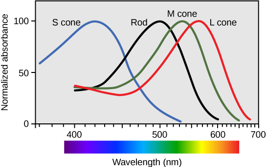
The longest wavelengths (lowest energies) in the visible spectrum are encoded by the visual system as red. The shortest wavelengths (highest energies) in the visible spectrum are encoded by the visual system as violet. In between lies an entire (literal) rainbow of colors: red, orange, yellow, blue, indigo, and violet. My engineer father taught me the mnemonic for this sequence by jokingly claiming the “inventor” of light was ROY G. BIV. I believed him.
If the membrane stack which extends from the photoreceptor cell body has the same diameter throughout, we call these cells rod photoreceptors. Rods are mostly indifferent to the wavelength of the incoming photons and are mostly interested in the number of photons. Incredibly, a rod photoreceptor can detect as few as 10 photons if the eye is perfectly adapted. This is the number of photons that enter the eye from a barely-visible distant star on a moonless night in the middle of the desert.
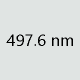 Rods are most sensitive to a single wavelength of light at about 500 nm (the actual number is a rather fussy 497.6 ± 3.3 nm) which appears to us as a gray-green color. As the wavelength increases above 500 nm or decreases below 500 nm, the rod photoreceptor becomes progressively less sensitive to light so it takes more photons to achieve the same electrochemical response. This is shown by the black curve.
If the membrane stack gets smaller as it gets further from the cell body, then we call this a cone photoreceptor. Cones are very sensitive to the wavelength of the incoming photon particle-waves. They code this information as color.
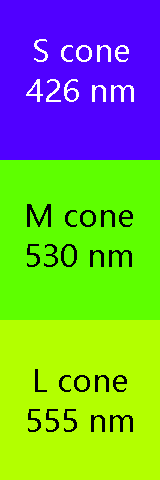 Cones come in three types. Long wavelength, or L cones, are sometimes called “red cones” though they respond best to a yellow-orange color at about 564 nm. Medium wavelength, or M cones, are sometimes called “green cones” and respond best to a yellow-green color at 534 nm. Short wavelength, or S cones, respond best to blue colors at 420 nm. (Note that the names are not the actual colors of the cones, but the colors that supposedly produce the “best” response.) Like the rods, each has a “favorite” color or wavelength. Photons with wavelengths near the “favorite” will cause a maximum response in that cone; as the wavelength moves away from the “favorite”, it takes more photons to produce the same response.
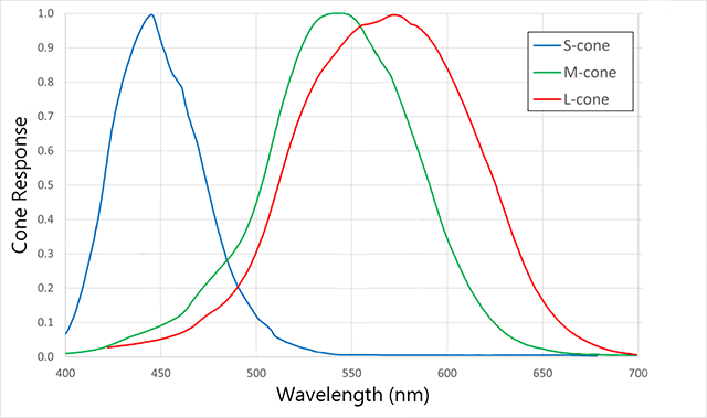
This setup allows the human retina to detect a complete range of colors in most cases. Each color in the visible spectrum gives us an unambiguous “readout” from the three cones. For example, using the diagram, note that a wavelength of 520 nm (green light) reads near zero on the S cone, at 60% on the L cone, and at 80% on the M cone.
A wavelength of 630 nm (red light) reads near zero on the S cone, at 60% on the L cone, and at about 10% on the M cone.
“Color Blindness” Is Actually Color Confusion
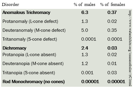 A problem arises if either the L cone pigment or the M cone pigment are missing or altered, which happens in about 9% of males. Then it is easy to confuse the color red with the color green, because both produce the same readout on the L cone if the M cone pigment is missing, or the same readout on the M cone if the L cone pigment is missing.
This condition is commonly called “color blindness” although people are not “blind” to these colors, they just confuse them. Because the genes for the M cone and L cone pigments are carried on the X chromosome, and women have two copies of this chromosome while men have only one, this relatively common mutation affects many more men than it does women.
Color Vision in Dogs
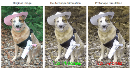
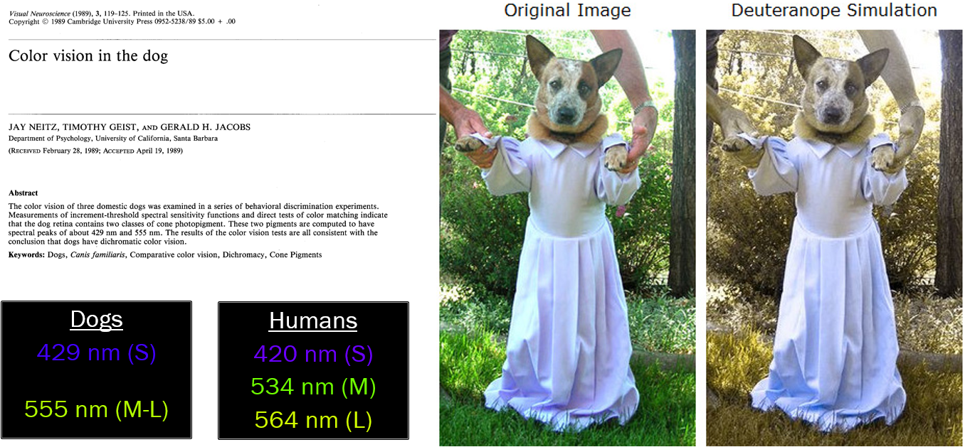
Like some humans, dogs and other mammals such as deer have only two cone pigments (with absorbance maxima at 429 nm or blue and 555 nm at yellow-green) and are prone to confuse red and green. That’s why deer cannot easily distinguish between hunter orange and foliage green and why dogs cannot easily distinguish an orange training dummy in green grass.
In the human retina, the other four cell types are “scraped away” at the foveal pit, leaving only the cone photoreceptors to detect the presence and wavelength of light. Thus the fovea (macula) is where visual acuity is the greatest. Almost all visual information comes from the central 5° of visual angle. (For comparison, the moon is about 0.5° of visual angle and the thumb at arm’s length is about 2° of visual angle, so the macula is about two and a half thumbs wide.) It takes a lot of motion, or a very large object, for us to detect visual stimuli outside of this central area. Without realizing it consciously, we move our eyes around to pick up elements of a visual scene in small ten-moon-diameter circles.
Just outside the macula, the number of cones decreases and rods predominate. This is why, to visualize a dim star, we need to look slightly away from it.

About 15° to the nasal side of the fovea is the blind spot, formed by the exiting ganglion cell axons that will form the optic nerve. Since no photoreceptors exist here, there is no perception of light from this region of the retina. Our visual system does a lot of work to make us unaware of the blind spot; contrariwise, we have to do a lot of work to be able to find our blind spot.
Media Attributions
- Trichromaticity © CNX OpenStax is licensed under a CC BY (Attribution) license
- 500 nm © Jim Hutchins is licensed under a CC BY-SA (Attribution ShareAlike) license
- Trichromaticity theory © Curran919 is licensed under a CC BY-SA (Attribution ShareAlike) license
- Cone absorbance maxima color blocks © Jim Hutchins is licensed under a CC BY-SA (Attribution ShareAlike) license
- Incidence of color blindness © Jim Hutchins is licensed under a CC BY-SA (Attribution ShareAlike) license
- Roxy deuteranope and protanope © Jim Hutchins & Hit N Heel No Red Light RA PT NA NAJ ("Roxy") is licensed under a CC BY-SA (Attribution ShareAlike) license
- Roxy deuteranope © Jim Hutchins is licensed under a CC BY-SA (Attribution ShareAlike) license
- Blind spot © Eric Chudler is licensed under a All Rights Reserved license
