Alpha Motor Neurons and the Neuromuscular Junction
Caleb Bevan
Objective 3: Describe α motor neurons (alpha motor neurons; lower motor neurons) and the nature of their synaptic contact with skeletal muscle.
α Motor Neurons
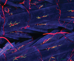
Motor neurons are stained red, muscles blue, and neuromuscular junctions are green and yellow in this immunofluorescence micrograph of Drosophila flight muscles. The neuron which controls the contraction of skeletal muscle is called an α motor neuron. Alternative names for this are alpha motor neuron (spelling out the Greek letter α). Clinicians call this the lower motor neuron to contrast them with the upper motor neuron. This is an important clinical distinction because the symptoms of lower motor neuron damage are completely different than the symptoms of upper motor neuron damage.
α motor neurons have their cell bodies in the ventral horn gray matter of the spinal cord. In humans, this area is called the anterior horn. They send axons out through a spinal nerve or cranial nerve, then end in an axon terminal with a presynaptic ending that contacts a skeletal muscle cell at a structure called the neuromuscular junction. There, the neurotransmitter acetylcholine is released, and diffuses across the synaptic cleft to bind to nicotinic acetylcholine receptors found on the postsynaptic membrane, in this case a skeletal muscle cell.
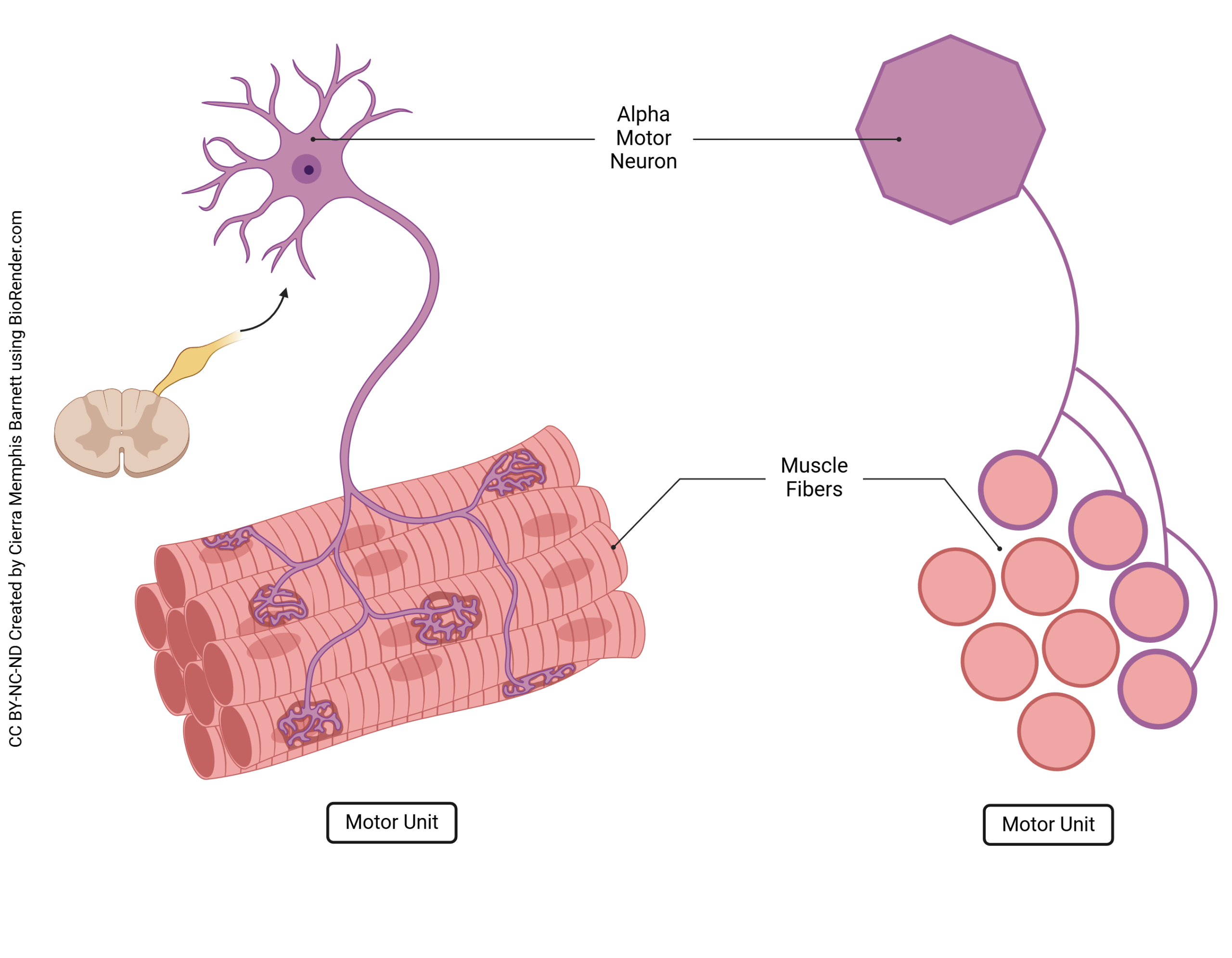
Each α motor neuron innervates between approximately 3 and 3000 muscle fibers. The population of muscle fibers innervated by a single α motor neuron is called the motor unit. A small motor unit size corresponds to fine control of muscle, while a large motor unit corresponds to the ability to generate a large amount of force from a single action potential in a single α motor neuron.
The Neuromuscular Junction
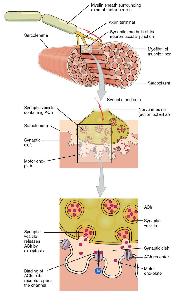
This image shows a series of magnifications of the neuromuscular junction. At top, a myelinated axon, about 10 μm in diameter, contacts a muscle fiber. As the action potential reaches the axon terminal, it causes the fusion of synaptic vesicles with the presynaptic membrane. This releases the neurotransmitter acetylcholine (ACh) into the synaptic cleft. ACh diffuses across the synaptic cleft and binds to nicotinic acetylcholine receptors embedded in the muscle cell membrane (sarcolemma). This opens a ligand-gated channel which allows the flow of Na+ into the cell, and also allows K+ to flow out of the cell. A small amount of Ca2+ can also travel into the muscle through this channel, but it is not physiologically significant. If one plugs an equal sodium and potassium permeability into the Goldman-Hodgkin-Katz equation, the calculated membrane voltage will be well above the threshold for triggering an action potential. This action potential has the same waveform as the action potential of an unmyelinated nerve axon.
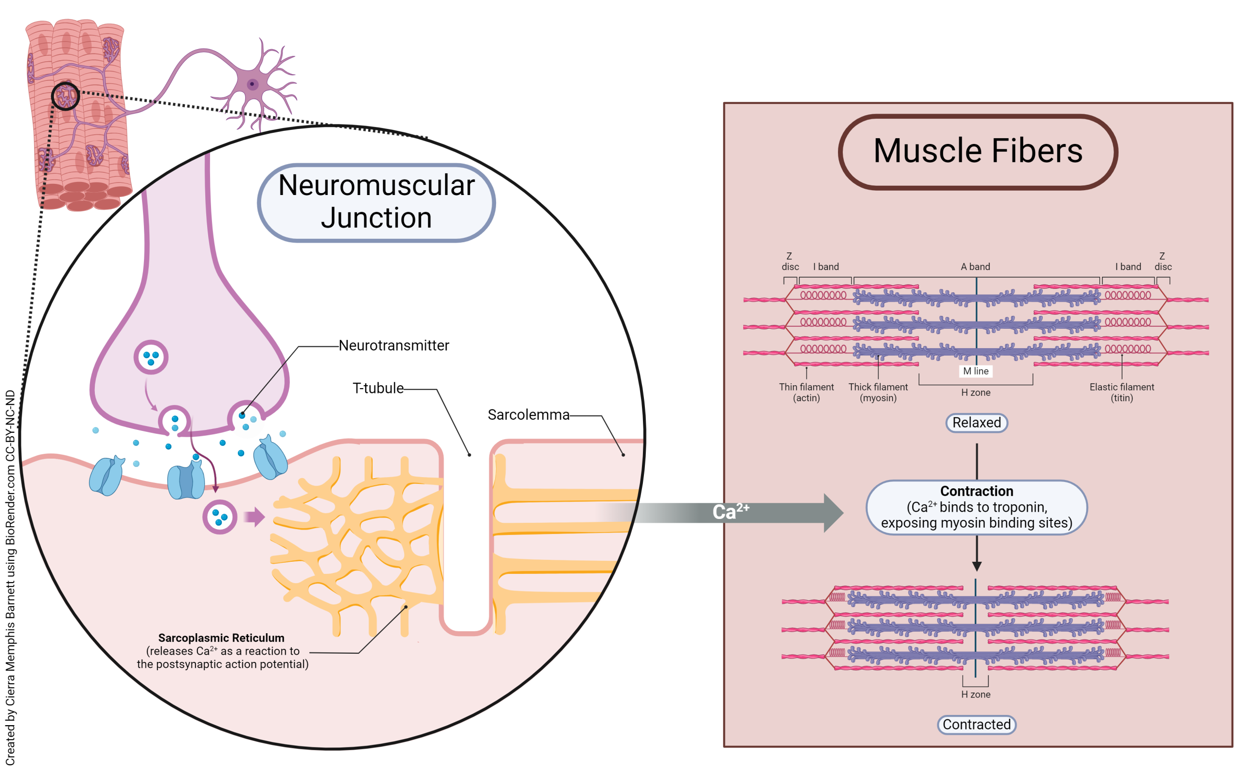
As we will see later, the muscle cell has extensive intracellular stores of Ca2+ sequestered within the sarcoplasmic reticulum, a type of smooth endoplasmic reticulum found only in muscle cells. The passing action potential opens voltage-gated Ca2+ channels and Ca2+ is released into the cytoplasm, where it can interact with regulatory proteins as we will see in the next chapter.
Upper Motor Neurons
All the other neurons in the brain and spinal cord that influence movement, but do not make direct contact with a skeletal muscle fiber, are called upper motor neurons by clinicians. Examples of these upper motor neurons are neurons of the precentral gyrus (primary motor cortex) of the brain; neurons in the basal ganglia (basal nuclei); and neurons of the cerebellum.
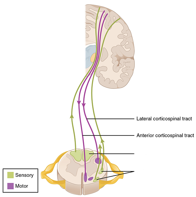
For example, neurons which make up the pathway from Brodmann area 4 (primary motor cortex) to the cell bodies of α motor neurons in the anterior horn of spinal cord are upper motor neurons. We will see this pathway in detail later in this section. This pathway includes pyramidal-shaped cell bodies in layer 5 of precentral gyrus; axons descending in the corona radiata, brainstem, and spinal cord; and axon terminals onto the α motor neuron in the ventral horn of spinal cord. This pathway is named the corticospinal (or pyramidal) tract and is one example of a set of upper motor neurons.
Media Attributions
- Neuromuscular junction © YJ Kim and M Serpe, NICHD is licensed under a CC BY (Attribution) license
- Motor Unit © Cierra Memphis Barnett is licensed under a CC BY-NC-ND (Attribution NonCommercial NoDerivatives) license
- Motor end plate © Betts, J. Gordon; Young, Kelly A.; Wise, James A.; Johnson, Eddie; Poe, Brandon; Kruse, Dean H. Korol, Oksana; Johnson, Jody E.; Womble, Mark & DeSaix, Peter adapted by Jim Hutchins is licensed under a CC BY (Attribution) license
- Muscle Fiber Innervation © Cierra Memphis Barnett is licensed under a CC BY-NC-ND (Attribution NonCommercial NoDerivatives) license
- Spinal cord pathways motor focus © Betts, J. Gordon; Young, Kelly A.; Wise, James A.; Johnson, Eddie; Poe, Brandon; Kruse, Dean H. Korol, Oksana; Johnson, Jody E.; Womble, Mark & DeSaix, Peter adapted by Jim Hutchins is licensed under a CC BY (Attribution) license
