Motion Processing in the Retina: Amacrine Cell and Bipolar Cell Inputs to Ganglion Cells
Jim Hutchins
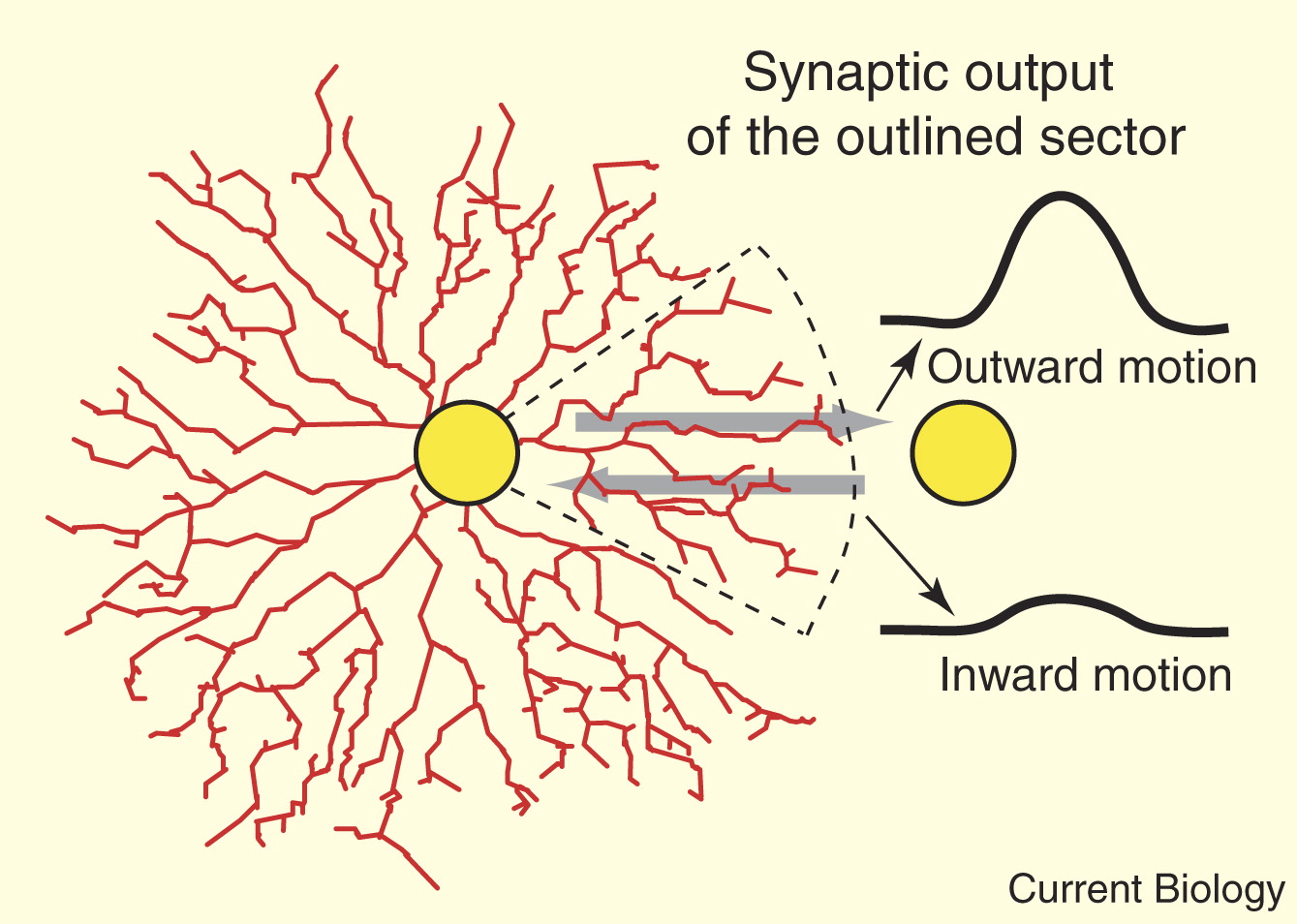
Objective 7: Summarize how amacrine cell and bipolar cell inputs to ganglion cells mediate motion processing.

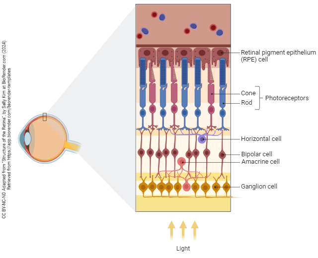
The cell bodies of horizontal cells and bipolar cells lie in a layer called the inner nuclear layer (INL). At the edge of the INL, another group of cell bodies are found; these belong to the amacrine cells. (The name “amacrine” means “axonless”; these cells, like horizontal cells, are all dendrites and no axon because they don’t send information over long distances.)
 Horizontal cells report on what nearby photoreceptors are doing. Amacrine cells report on what nearby bipolar cells are doing. This means that if an edge is moving, it’s the amacrine cells that make that report. If an edge is change, then they report on change in change. (In mathematical terms, that’s the second derivative, but if those words mean nothing to you, then ignore them.)
Bipolar cell terminals and amacrine cell terminals synapse onto ganglion cell dendrites in the inner plexiform layer (IPL), also called the inner synaptic layer. Amacrine cell bodies are also found lining the other edge of the IPL in the misnamed ganglion cell layer (GCL).
Media Attributions
- Starburst amacrine cells fried and masland © Shelley Fried and Richard Masland is licensed under a All Rights Reserved license
- Structure of the retina cell types © Sally Kim adapted by Jim Hutchins is licensed under a CC BY-NC-ND (Attribution NonCommercial NoDerivatives) license

