Types of Muscle Tissue
Objective 10.2
10.2.1 Identify and name important features of the three types of muscle tissue: skeletal, cardiac, and smooth muscle.
10.2.2 Compare and contrast the three different types, paying attention to the appearance of the cell, the arrangement of the cells and the basic functions for each type.
A common theme in anatomy and physiology is that form and function are closely related. This is especially true for the types of muscles. What are the similarities between these classes of muscles? What are the major differences? How do these similarities and differences allow these muscles to perform their unique functions?
Skeletal muscle is named such because of its connection to the skeleton. This relationship allows contractions of the muscles to move the body or maintain posture. Skeletal muscle is under voluntary control, meaning it requires conscious thought and intentional stimuli from the nervous system in order to contract. You have more than likely seen what happens when an individual loses consciousness; their control over skeletal muscle immediately ceases.
The movement of our skeletal muscles is the job of many different areas of the brain, but we will wait until a discussion of the nervous system to tackle that. Generally speaking, these kinds of movements are referred to as motor movement. The neurons of the nervous system that cause these movements, coincidentally, are called motor neurons. Upper motor neurons are those that originate in the brain as well as those that send signals within the brain and down the spinal cord. Lower motor neurons extend from the spinal cord to the muscle or group of muscles they innervate.
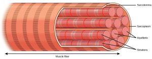
A key feature of skeletal muscle cells is the arrangement of the contractile proteins actin and myosin. The highly organized structure is called a sarcomere. Sarcomeres are arranged end to end in a repeating fashion throughout the muscle. This gives the appearance of striations (stripes). The sarcomere is the basic unit of muscle contraction. One sarcomere is about 2.5 μm long. When the sarcomeres collapse on themselves and become shorter, the entire muscle becomes shorter. The large rectus femoris muscle of the leg contains perhaps 150,000 sarcomeres end to end; as each of these shortens by a tiny amount, the entire muscle is contracted.
In skeletal muscle, thousands of sarcomeres, arranged end to end, form long tubes called myofibrils. These tubes are wrapped together in connective tissue and make up the bulk of the skeletal muscle cell (muscle fiber). Due to the length of each muscle fiber, many nuclei are scattered along the length of the cell. The average skeletal muscle cell is about 3 cm in length, but they can vary from about 1 mm (stapedius muscle of the middle ear) to over 50 cm (sartorius muscle of the leg).
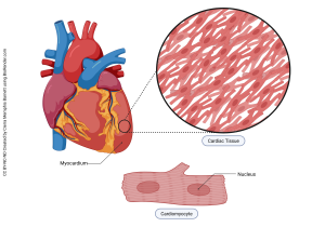
Cardiac muscle has a similar appearance to skeletal muscle because it is comprised of sarcomeres, however each cardiac muscle cell displays a branching appearance instead of the long, narrow tube-like appearance of skeletal muscle cells. The purpose of cardiac muscle contraction is to squeeze the heart and push blood out of the chambers and into the arteries, capillaries, and veins of the body with just enough force to return it back to the heart (Unit 16). A branching arrangement of cells allows for this type of movement. Like skeletal muscle, the coordinated shortening of thousands of sarcomeres results in the contraction of the heart muscle.
Cardiac muscle is unique in that it responds to signals that arise from specialized cells within the heart itself. These cells send electrical signals about every 0.8 seconds, causing the heart to contract about 72 times per minute (called heart rate or pulse). Under stress, hormones, nervous system signals, or both can cause the heart rate to increase. Similarly, signals from the body can slow the heart rate during periods of rest. The constant rate of contraction as well as the feedback loops involved in increasing or decreasing the heart rate are involuntarily controlled — lucky for us. Can you imagine having to consciously tell your heart to beat 72 times a minute? The penalty for forgetfulness would be severe.
With skeletal muscle, it is possible to contract a few individual fibers or entire groups of muscles all at once. With cardiac muscle, it is critical that all of the muscle fibers contract in unison. In order to achieve this, the heart muscle cells have a series of gap junctions that transmit the electrical signal for contraction all the way through the entire muscle simultaneously.
Structurally, the cardiac muscle needs to be strong so that it doesn’t rip itself apart with the constant, regular contractions. In order to do this, the heart has intercalated discs seen as zigzag lines on the surface of the drawing of cardiac cells above. These intercalated discs are regions with multiple desmosomes holding the muscle cells together. Remember that desmosomes are “spot-welds” that give a tissue structural integrity and strength.
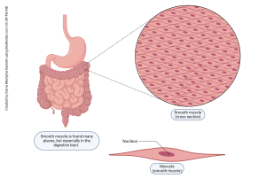
Smooth muscle gets its name because it lacks the sarcomeres that give skeletal and cardiac muscle their striated appearance. Smooth muscle still makes use of actin and myosin proteins, but the arrangement appears more unorganized than in the other muscle types. The seemingly random arrangement of contractile proteins results in more of a squeeze-type contraction. This type of is more appropriate for moving substances throughout the digestive system or reducing the diameter of a blood vessel or the pupils of the eye. Smooth muscle contractions are typically slower to initiate, but last longer than other muscle types.
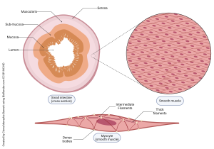
Smooth muscle is located in the walls of hollow internal structures, such as blood vessels, airways, lining the digestive system and in most organs of the abdominal cavity. It is also seen in other areas like the skin, eyes and reproductive organs.
Smooth muscle is not under voluntary control. Blood vessels expand and contract, airways open and constrict, our pupils dilate and constrict, and our gut keeps things moving, even while we sleep or while we’re otherwise distracted with other things.
The nerves that innervate smooth muscle are there to regulate smooth muscle contraction rather than control it. That is, the nerve cells release chemicals that increase or decrease the overall activity level of smooth muscle, but they don’t control individual muscle cells as much as in skeletal muscle.
The digestive system has its own nervous system that helps coordinate and control contraction of large blocks of smooth muscle, to keep things moving from mouth to anus without needing the brain to tell it what to do. The brain is capable of making helpful suggestions, but it doesn’t run the show in smooth muscle.
Media Attributions
- U10-008a skeletal muscle cell © Betts, J. Gordon; Young, Kelly A.; Wise, James A.; Johnson, Eddie; Poe, Brandon; Kruse, Dean H. Korol, Oksana; Johnson, Jody E.; Womble, Mark & DeSaix, Peter is licensed under a CC BY (Attribution) license
- U10-008 Figure_38_04_02 © Clark, Mary Ann; Douglas, Matthew; Choi, Jung is licensed under a CC BY (Attribution) license
- U10-007 Cardiac Muscle © Barnett, Cierra Memphis is licensed under a CC BY-NC-ND (Attribution NonCommercial NoDerivatives) license
- U10-010 Small Intestine Smooth Muscle © Barnett, Cierra Memphis is licensed under a CC BY-NC-ND (Attribution NonCommercial NoDerivatives) license
- U10-011 Smooth Muscle © Barnett, Cierra Memphis is licensed under a CC BY-NC-ND (Attribution NonCommercial NoDerivatives) license
