The Plasma Membrane
Objective 4.3
4.3.1 List and classify the components of the plasma membrane, as defined by the fluid-mosaic model.
4.3.2 Classify membrane proteins as peripheral or integral.
4.3.3 Define permeability.
4.3.4 Classify substances according to permeability through the plasma membrane.
In 1973, Singer and Nicholson proposed a new model for the cell membrane, which came to be called the fluid-mosaic model. Among the features of the model are:
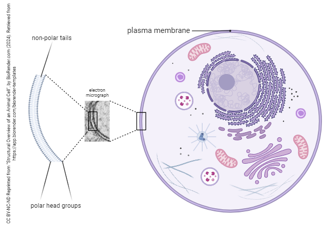
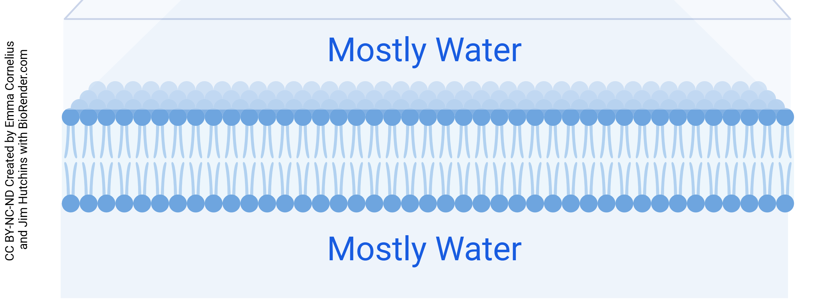
- Lipids consist of polar (i.e. hydrophilic, water-soluble) head groups with non-polar (i.e. hydrophobic, fat-soluble) tails.
- Lipids arrange themselves into two layers, with the head groups facing the extracellular and intracellular solutions (like the “bread” in a sandwich) and with the lipid tails forming a layer between (like the “peanut butter” of a sandwich).
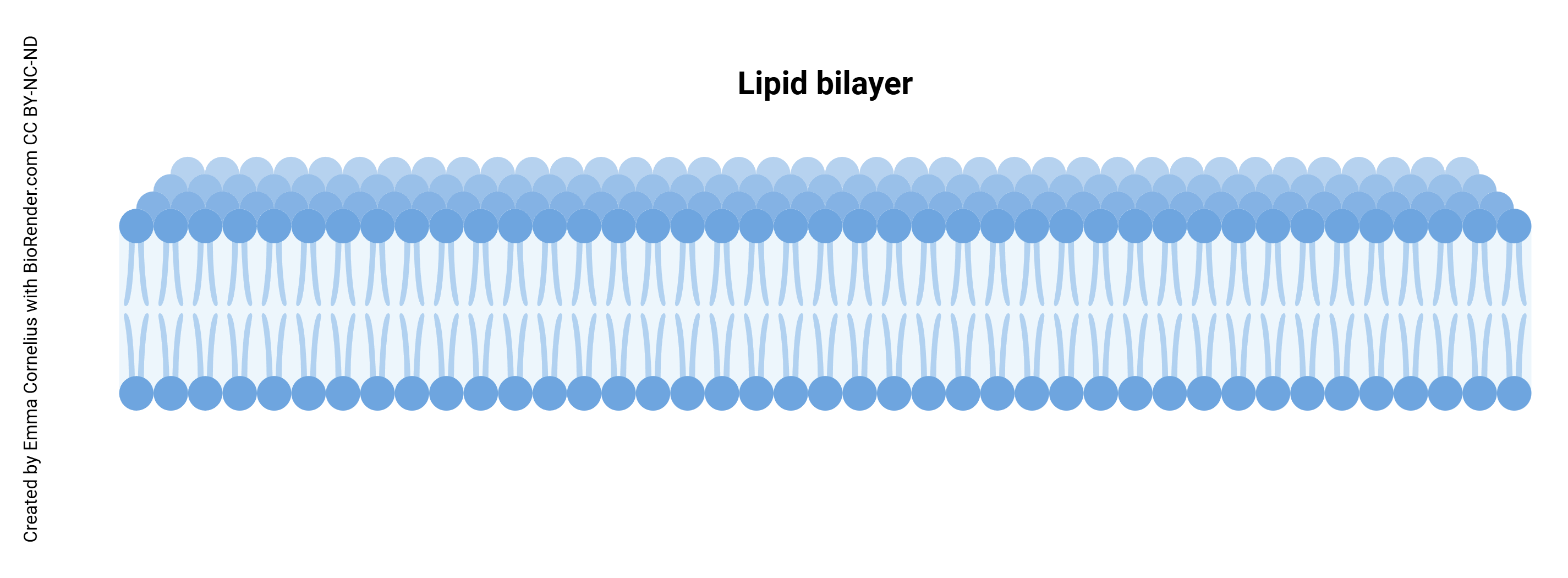
- These lipids can move side-to-side, but never flip from the outside to the inside or vice-versa.
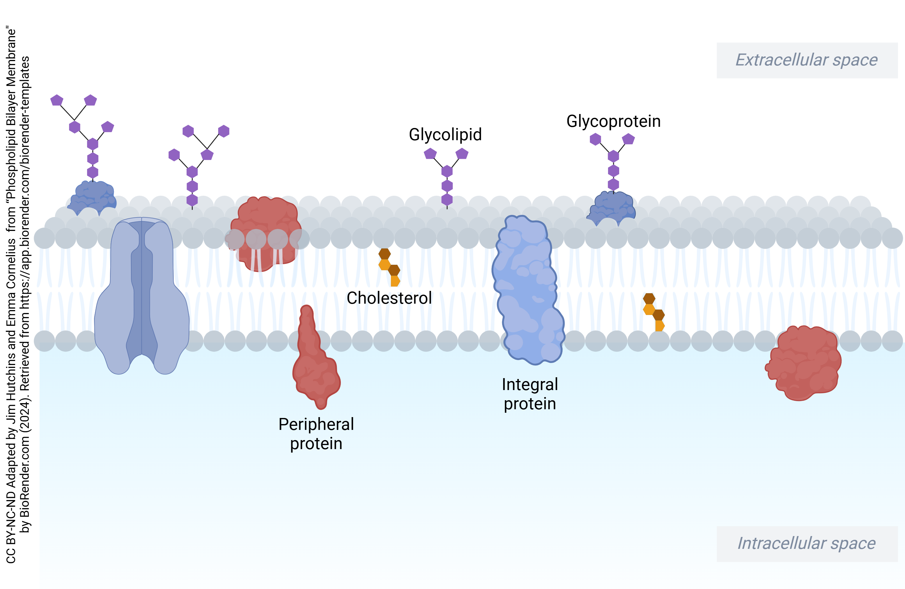
- Proteins are embedded in the lipid “sea” and are free to float around side-to-side, but like the lipids, these never flip over (to do so would expose the hydrophilic regions to the hydrophobic core of the membrane, a huge no-no).
- Protein arrangements can be in one of two broad categories: peripheral proteins that associate with either the extra or intracellular lipid heads, or integral proteins that span the membrane (transmembrane proteins) with hydrophobic amino acid residues hanging out with the lipid tails in the hydrophobic core of the sandwich.
plasma membrane = cell membrane = plasmalemma
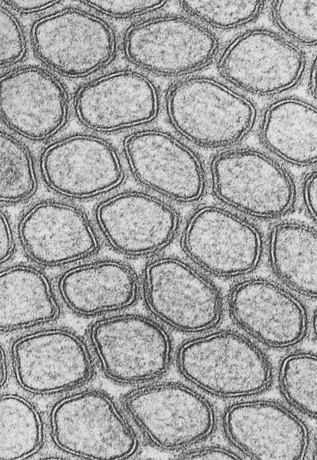
Using the electron microscope, we can see the plasma membrane (PM) as two black lines that always run parallel to each other. These two parallel lines are the polar head groups which bind to osmium atoms, which are used as a stain in electron microscopy.
Peripheral membrane proteins are loosely associated with the cell membrane and lie either completely on the outside or completely on the inside of the cell. On the other hand, integral (transmembrane) proteins span the cell membrane from outside to inside the cell.
A “naked” lipid bilayer without proteins allows water across the cell membrane, but the reasons are a mystery to almost everyone who studies membranes. It doesn’t matter for “real” cells, because scientists estimate that in red blood cells, for example, 90% of the water that passes through the membrane goes through a water pore protein called an aquaporin2. It appears that most (if not all) body cells contain enough aquaporin to overwhelm any resistance to water passing through the membrane. Aquaporin is important for kidney function, as we will see in a later module.
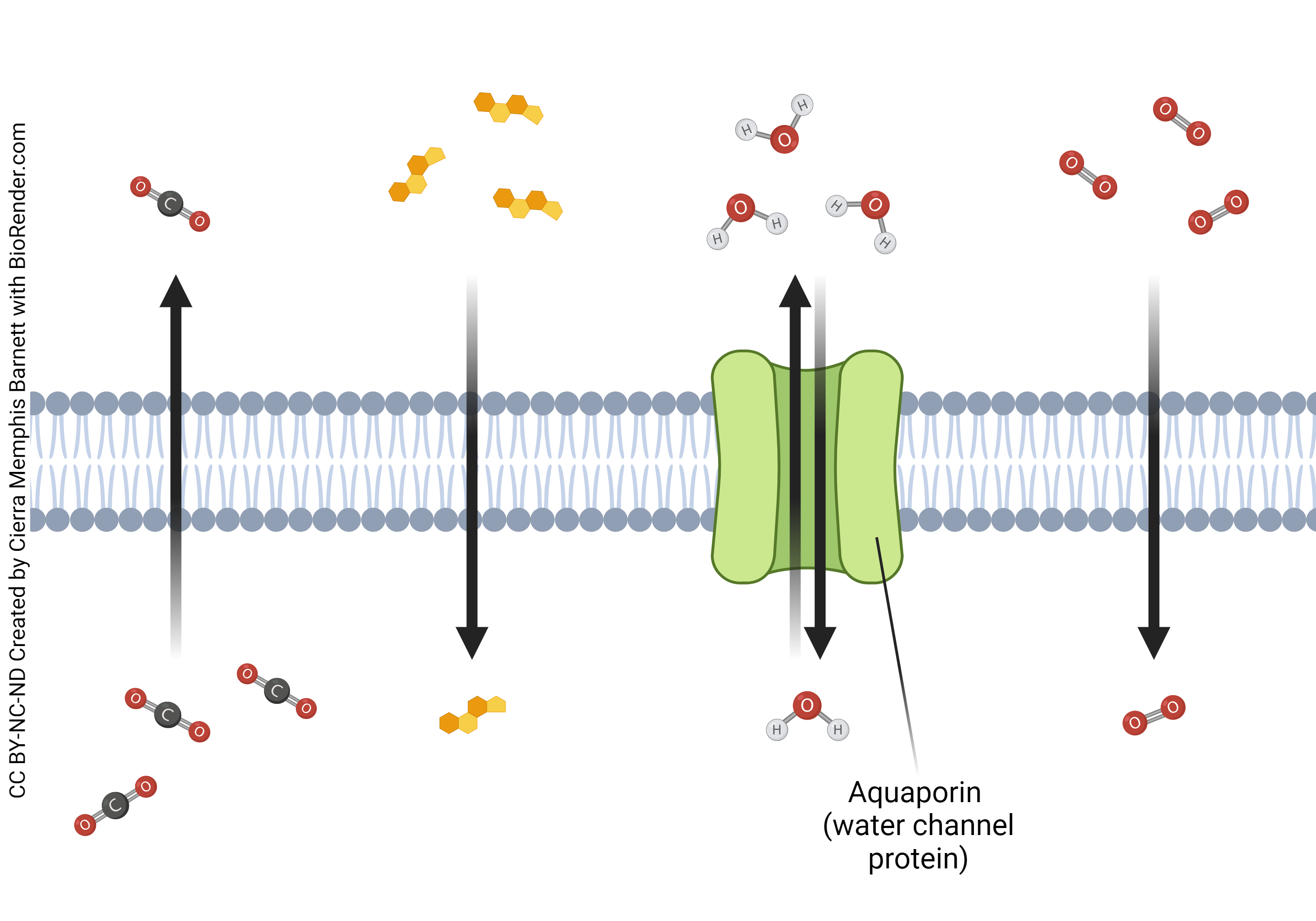
Gases pass easily through the lipid bilayer, because a gas molecule is small and non-charged Similarly, small, fat-soluble molecules such as butane and propane pass easily. That’s why these solvents are popular with young people who dissolve the lipid bilayer in their brains by “huffing”. There are no old people who huff, which should tell you something. Charged ions and large molecules don’t pass through the lipid bilayer without assistance.
Water is found on both sides of the cell membrane. Extracellular fluid is found outside of the cell, intracellular fluid is found inside of the cell. The polar heads of the phospholipids are hydrophilic, water loving. Because of this, they are oriented towards the outside or inside of the cell. Charged ions and water-soluble substances are attracted to the hydrophilic heads and will hang out there, rather than cross the membrane. Just like you might not want to swim across the English Channel, water-soluble substances need a bridge. We’ll talk more about this when we cover membrane proteins.
Media Attributions
- U04-010 lipid bilayer © Cornelius, Emma is licensed under a CC BY-NC-ND (Attribution NonCommercial NoDerivatives) license
- U04-007 Structural Overview of an Animal Cell preview Unit 4 © BioRender is licensed under a CC BY-NC-ND (Attribution NonCommercial NoDerivatives) license
- U04-019 em microvilli FawcettTheCellChapter1 p8 © Fawcett, Don is licensed under a CC BY-NC-ND (Attribution NonCommercial NoDerivatives) license
- U04-012 and 029 phospholipid bilayer © Hutchins, Jim & Cornelius, Emma is licensed under a CC BY-NC-ND (Attribution NonCommercial NoDerivatives) license
- U04-021 same as U13-001 Simple Diffusion © Barnett, Cierra Memphis is licensed under a CC BY-NC-ND (Attribution NonCommercial NoDerivatives) license
- U04-022 lipid bilayer with water © Cornelius, Emma and Hutchins, Jim is licensed under a CC BY-NC-ND (Attribution NonCommercial NoDerivatives) license
