The Neuromuscular Junction
Objective 10.5
10.5.1 Characterize the neuromuscular junction.
10.5.2 Label the components of the neuromuscular junction on a diagram and explain their role in triggering a muscle contraction.
10.5.3 Correlate the electrical signal from the nervous system and the twitch or contraction from the muscle.
Skeletal muscle is controlled using conscious signals generated by the nervous system (Unit 12). In this objective and the next, we will study excitation-contraction coupling, how nerve and muscle cell excitation or electrical activity is turned into the shortening of sarcomeres.
We will see in Unit 13 that a lipid-rich wrapping called myelin is electrical insulation found on nerve axons (“wires”). The nerve cell axon terminates at an axon terminal. These axon terminals are bulb-shaped, and they are called by several names, but for now let’s call them synaptic end bulbs.
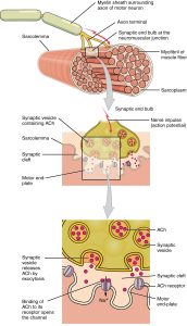
Inside the synaptic end bulb are vesicles (Latin: “little bladders”) filled with a signaling molecule, acetylcholine (ACh). There is a gap between the motor neuron synaptic end bulb and the muscle cell receptive surface. This gap is called the synaptic cleft. The receptive surface of muscle is called the motor end-plate.
Through the process of exocytosis, which we first studied in Unit 4, ACh is released into the synaptic cleft. It moves across the cleft by diffusion. It will blindly encounter an acetylcholine receptor protein (Unit 4) on the receptive surface of the motor end-plate, and cause excitation in the muscle by movement of ions (Unit 2).
An electrical impulse (red lightning bolt) arrives at the synaptic end bulbs of the motor neuron.
As we will see in detail in Unit 13, the electrical impulse results in the exocytosis of the signaling molecule acetylcholine (ACh). ACh diffuses across the synaptic cleft.
ACh receptors at the motor end-plate bind ACh and open an ion channel which allows the charged ion sodium (Na+), and other charged ions not shown, to enter the muscle cell and cause it to depolarize (i.e. become more positive). This excites the muscle cell.
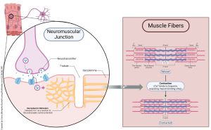
When the motor neuron receives a proper stimulus, it releases acetylcholine across the synapse and onto the sarcolemma of the muscle fiber. There, specialized receptors turn the chemical signal into an electrical signal. As long as ACh is present in the synapse, the action potential will be generated in the muscle. When the neuron is no longer sending the signal, the ACh is removed from the synapse by an enzyme called acetylcholinesterase and the muscle relaxes until the next signal arrives.
The electrical signal from the ACh receptor spreads over the entire surface of the muscle fiber, penetrating into the T tubules (transverse tubules) where they meet the sarcoplasmic reticulum (SR) at triads. This causes calcium release from its stores in the SR, which begins the contraction phase of muscle contraction coupling.
As mentioned previously, both nerve and muscle tissues are called excitable tissues. This means they send signals through changes in their electrical properties, especially a change in voltage (electrical potential). In many neurons and all skeletal and cardiac muscle, this electrical change is called an action potential. That is, it is an active change in the electrical potential (voltage) of the neuron and muscle cell.
A single motor neuron branches several times and contacts muscle fibers. One neuron can make contact with a varying number of muscle fibers, from three up to three thousand. The neuron that stimulates the muscle fibers and the fibers that respond are collectively referred to as the motor unit. When we make very small, precise movements, like with our eyes, vocal cords, or fingers, we stimulate only a few muscle fibers at a time.
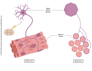
- A single neuron and all the muscle fibers it inntervates are a motor unit
- The motor unit is the smallest division that the system can control individually
These motor units would be considered very small. Movements that require more muscles or greater force, like postural muscles, or walking/running muscles, have larger motor units. Large motor units stimulate large groups of muscles at a time, sometimes stimulating thousands of fibers with only one motor neuron. Different motor units are used for different types of movements depending on the amount of strength and precision required.
“Nerve impulse” = action potential
All of the reactions we just discussed happen very quickly, typically only milliseconds at a time. So how then can you contract a muscle and hold the contraction for extended periods of time? When muscles are stimulated by neurons, they demonstrate a brief lag known as a latent period. The muscle then contracts and immediately relaxes, all happening within a period of about 50 milliseconds. In order to sustain muscle contraction, the nervous system continuously sends electrical impulses. When these signals come frequently enough to limit the time the muscle fibers have to relax, it is referred to as wave summation. Wave summation is responsible for the strength that comes from muscles that contract without relaxing. If you are sitting there reading this, you should be grateful for wave summation and the way it is helping you to maintain your current posture. If you are laying and reading this, how are you still awake?
Media Attributions
- U10-023 1009_Motor_End_Plate_and_Innervation-1 © Betts, J. Gordon; Young, Kelly A.; Wise, James A.; Johnson, Eddie; Poe, Brandon; Kruse, Dean H. Korol, Oksana; Johnson, Jody E.; Womble, Mark & DeSaix, Peter is licensed under a CC BY (Attribution) license
- U10-025 Muscle Fiber Innervation © Barnett, Cierra Memphis is licensed under a CC BY-NC-ND (Attribution NonCommercial NoDerivatives) license
- U10-024 Motor Unit copy © Barnett, Cierra Memphis is licensed under a CC BY-NC-ND (Attribution NonCommercial NoDerivatives) license
