The Cell Cycle
Objective 6.6
6.6.1 Describe the stages of the cell cycle.
6.6.2 Describe the events that take place in each stage.
6.6.3 Be able to recognize each stage in diagrams or photographs.
6.6.4 Know the functional significance of each stage.
Cells of the body renew themselves by cell division. Tissues that are constantly regenerating always undergo cell division: skin, bone marrow making red and white blood cells, and the intestinal lining.
There are also cells of the body that do not divide to any appreciable extent. It is difficult to prove that no cell division is taking place, but it can be demonstrated that cell division is difficult and slow in these tissues. Among the tissues in this category are: smooth muscle, skeletal muscle, heart (cardiac) muscle, and neurons (nerve cells) of the brain.
Cells which are dividing go through a series of steps called the cell cycle.
The cell cycle is stereotyped: all cells go through the same stages in the same order, and each stage takes about the same amount of time.
The actual process of cell division is called mitosis or M phase. Mitosis has its own stages that we will discuss later. Collectively, all the other phases — in order, G1, S, and G2 — are referred to as interphase.
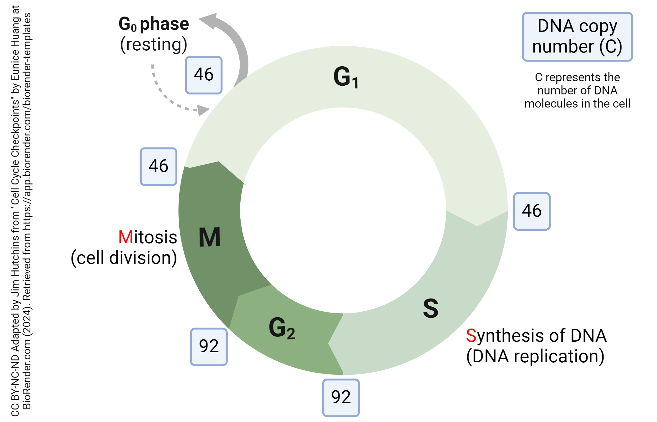
On the diagram shown here, we’ve also indicated the number of DNA molecules (white number) in the cell at the beginning of each stage of the cell cycle. Normal cells spend most of their life in a diploid state. There are two copies (C) of each DNA molecule (i.e. two of chromosome 1, two of chromosome 2, etc.) The diploid state is symbolized 2N.
Ploidy is a term that refers to the number of chromosomes; diploid cells have two copies each of the numbered chromosomes (autosomes) 1-22 (numbered in decreasing size order) for a total of 44 autosomal DNA molecules as well as the sex chromosomes (XX or XY); these two added to the 44 autosomes make a total of 44 + 2 = 46 DNA molecules in the diploid cell. Cells at the end of mitosis contain 46 DNA molecules (46C).
After mitosis, a dividing cell enters a first growth (or “gap”) phase called G1 (“Gee-one”). G1 takes about 10 hours to complete.
Next, the cell passes from G1 to S phase. “S” stands for “synthesis”, in this case, synthesis of DNA. During S phase, the cell’s DNA is copied (replicated) so that it can be divided equally between the daughter cells in mitosis. The number of DNA molecules doubles from 46 to 92. The cell at the end of S phase is now 92C.
This process takes about 8 hours.
After S phase, cells enter a second growth phase or second gap phase called G2. During G2, the cell finalizes its preparations for mitosis. After about 4 hours of G2, the cell enters M phase with 92 DNA molecules (92C).
In order to pass from one part of the cell cycle to another, the cell must pass through a checkpoint. These checkpoints are an important regulator of the cell cycle, and therefore control whether the cell divides or not. Cancer cells lose the ability to stop cells at checkpoints and the uncontrolled growth which results is an important part of the biology of cancer. If you’ve ever heard of someone having a 2 or 3 kg tumor removed, that was the result of uncontrolled cell division. (The Guinness World Record for tumor size is 139 kg.)
Probably the most important checkpoint in cell division is the restriction point. A cancer-causing virus, simian sarcoma virus, has a defect in this checkpoint which allows cells to cycle out of control. There are also DNA damage checkpoints during S and G2 phase which remove cells from the cycle if they have irreparable DNA damage.
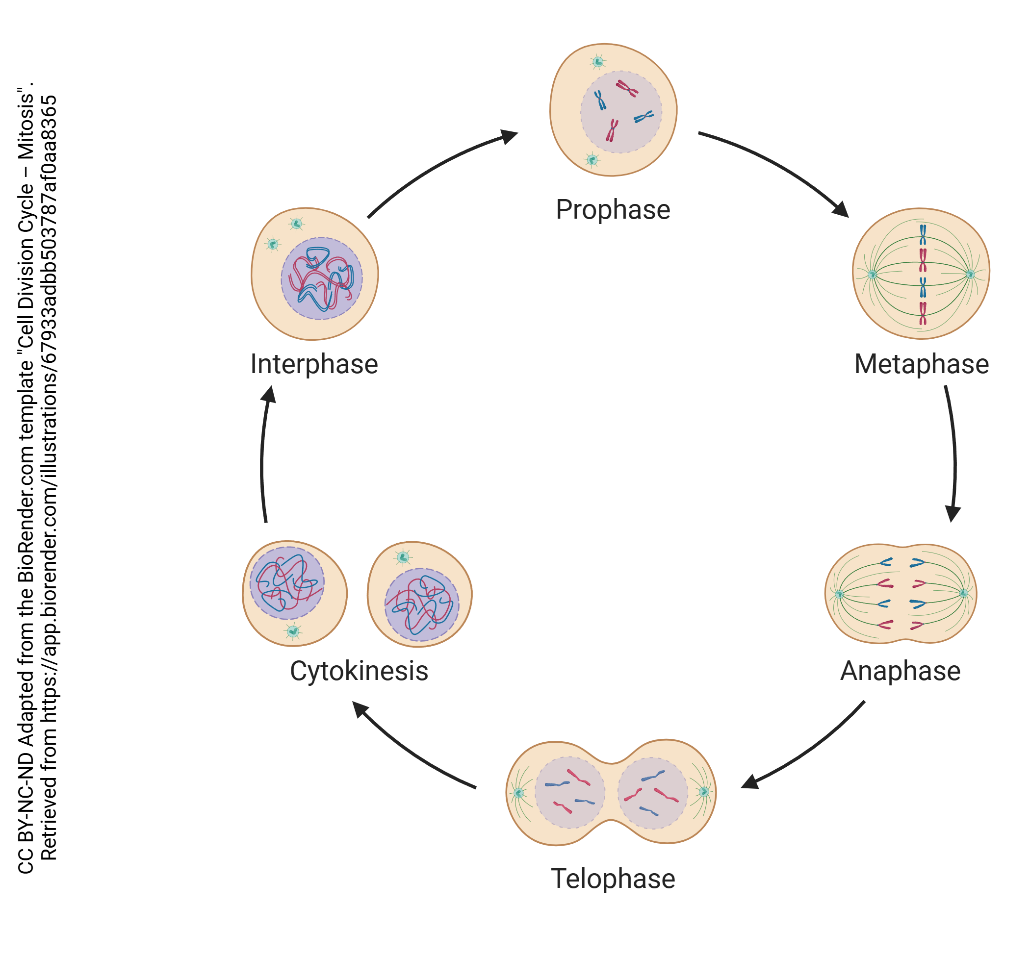
Mitosis has four parts: prophase, metaphase, anaphase, and telophase.
As the last step in mitosis, the cells must physically divide into two daughter cells. This physical division is called cytokinesis (cyto-, “cell”; -kinesis, “moving”).
While all parts of interphase involve preparation for, or recovery from, mitosis, the first phase of mitosis that involves an obvious, observable visual change in the cell is prophase. That change is the condensation of loose, unspooled chromatin into tightly-packed chromosomes that are visible under a light microscope.
Chromosomes are highly condensed DNA wound around histone proteins. (Greek khroma–, “color” + –soma, “body”, because these were colored bodies visualized through the light microscope with dyes used by German histologists of the late 19th century.)
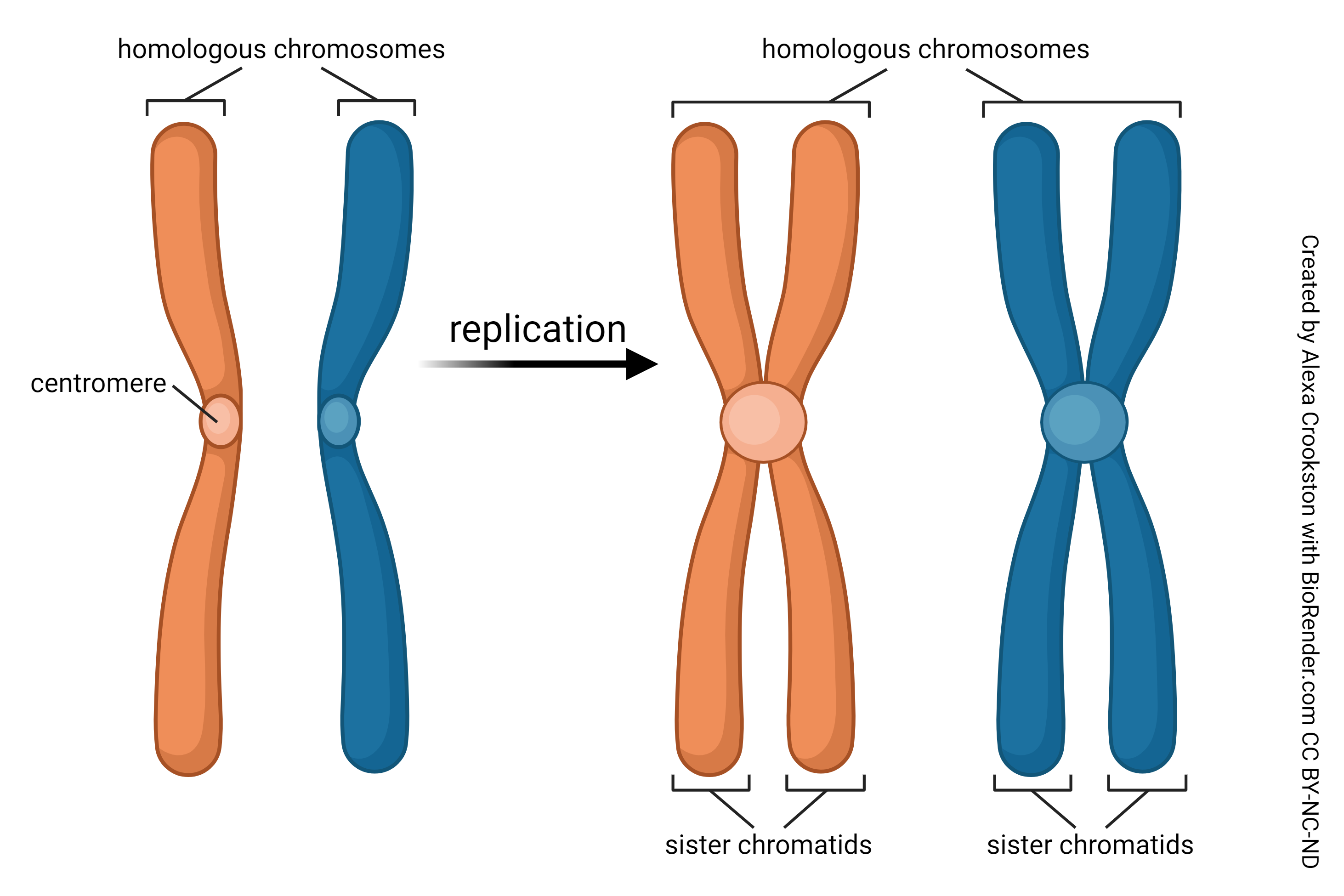
Eukaryotic chromosomes have a distinctive structure, and are more-or-less shaped like the letter “X”. The center of the X is called the centromere (Greek & English centro– + – meros, “part”). There is a shorter arm, named the p arm after the French petite (“little”) and a longer arm, named the q arm because who knows but probably it’s the letter after p in the alphabet. At the ends of the chromosome are regions that are the subject of intense interest by those scientists studying the biology of cancer, called telomeres (“end part”).
A chromosome consists of two chromatids. The two chromatids, joined at the centromere, are almost perfect copies of each other, with only slight variations that we will study later as Mendel’s alleles. Thus, there are two copies of chromosome 1, each with two chromatids for a total of four copies of the same gene sequences in prophase cells just before they divide. Females have two paired copies each of the 22 autosomes (44 numbered chromosomes, 88 chromatids or 88 DNA molecules) plus two paired copies of the X chromosome (XX, 4 chromatids or 4 DNA molecules). That means they have 92 (= 88 + 4) DNA molecules in each prophase cell. Males (XY) have a similar setup, with 88 DNA molecules representing autosomes, 2 chromatids or 2 DNA molecules making up the X chromosome, and 2 chromatids or 2 DNA molecules making up the Y chromosome for the same 92 DNA molecules in the prophase cell (= 88 + 2 + 2). That’s where we get the number “92” as shown on the cell cycle diagram shown at the beginning of this objective.
In prophase, DNA is tightly packed into chromosomes, the nuclear envelope breaks down, and the mitotic spindle forms.
The mitotic spindle, which grows out of the microtubule organizing center called a centrosome, is made up of the microtubules used to move chromosomes.
Metaphase, as the name implies, is the middle phase of mitosis, in which everything in the parent cell lines up in the middle.
Chromosomes, formed during prophase, move to the middle of the parent cell and the microtubules of the mitotic spindle attach to anchors (centromeres) found in the middle of each chromosome.
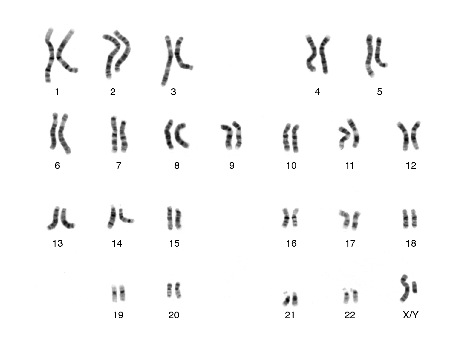
This is a picture of chromosomes called a karyotype. Notice 2 DNA strands make up each pair of chromosomes. One set of this DNA comes from mom, one from dad. There are 22 pair of autosomal chromosomes. The 23rd pair of chromosomes determine gender. Notice this individual has an X and Y chromosome which would denote this individual is biologically male. This cell would be diploid; there are 2 of each chromosome.
The prefix “ana–” means “backward”, so anaphase is the stage of mitosis where the contents of the two daughter cells move backwards away from each other.
The chromatids (single DNA molecules, forming the “arms” of chromosomes) are torn apart into two equal pieces by the action of the mitotic spindle. The chromatids then back up and take their places on either end of the elongating cell.
During telophase (Latin telo–, “end”), mitosis is completed. A cleavage furrow appears in the middle of the parent cell, and as the furrow deepens, the cell is split into two daughter cells.
In each of the daughter cells, the nuclear envelopes reform and the mitotic spindle comes apart.
The daughter cells move apart in a process called cytokinesis (“cell motion”).
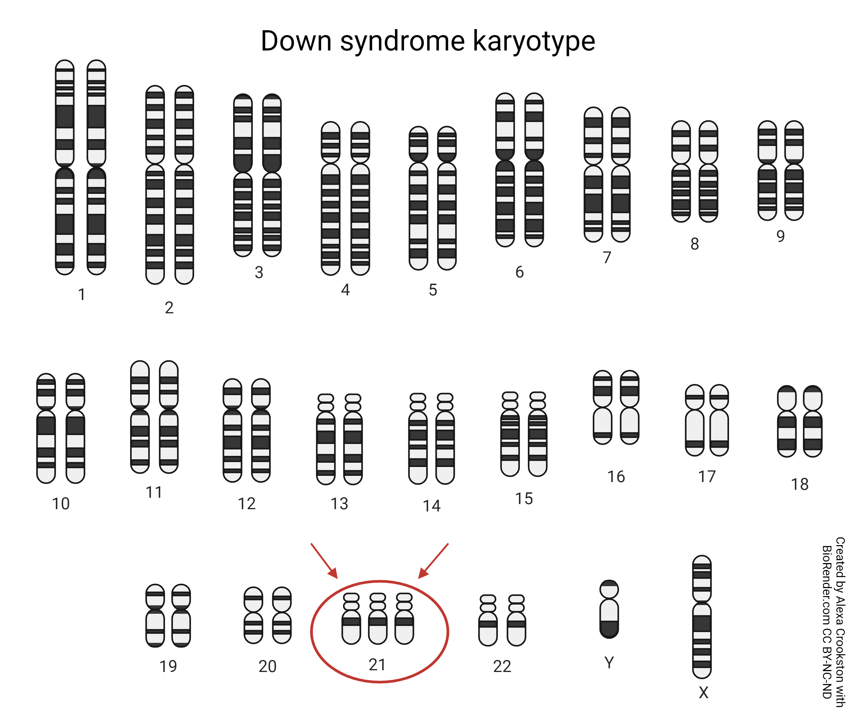
Metaphase is the stage of mitosis used to create a karyotype, a picture of the chromosomes that is used (for example) to diagnose disorders with a change in the number of chromosomes. Trisomy 21, or Down syndrome is an example.
Now, the two daughter cells have re-entered G1. The process of mitosis, which began with one cell, has now created two cells in interphase.
Media Attributions
- U06-035 same as U20-037B Cell Cycle Mitosis 2100px © Huang, Eunice adapted by Jim Hutchins is licensed under a CC BY-NC-ND (Attribution NonCommercial NoDerivatives) license
- U06-036 Cell Division Cycle – Mitosis © BioRender is licensed under a CC BY-NC-ND (Attribution NonCommercial NoDerivatives) license
- U06-037 Chromosome Replication © Crookston, Alexa is licensed under a CC BY-NC-ND (Attribution NonCommercial NoDerivatives) license
- U06-038 karyotype © National Human Genome Research Institute is licensed under a Public Domain license
- U06-039 Down Syndrome Karyotype © Crookston, Alexa is licensed under a CC BY-NC-ND (Attribution NonCommercial NoDerivatives) license
