Structural Organelles
Objective 5.10
5.10.1 Name and classify the organelles involved in cellular structural integrity.
5.10.2 Identify the structural proteins involved.
5.10.3 Describe how these proteins support microvilli, attachments between cells and cell division.
Cytoskeleton
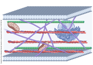
This drawing, modified from a pen and ink original by Keith Porter (who trained Jim Hutchins), shows the complicated geometry of the cytoskeleton as seen through the high voltage electron microscope.
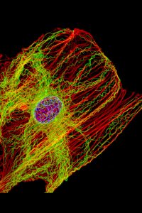
In the past 50 years since Fawcett made his pen-and-ink drawings, we’ve advanced microscopy to the point where we can visualize the cytoskeleton directly using fluorescence microscopy, as seen here.
The cytoskeleton (“cell skeleton”) is to the cell as our skeletal system is to our bodies. It is made of a number of different proteins that form thread- or tube-like structures. These proteins provide structural stability, move substances around in the cell, and either move the cell around in the extracellular environment, move the extracellular environment around the cell, or move the intracellular environment around inside the cell. They are classified, by size (largest to smallest), as microtubules, intermediate filaments, or microfilaments (actin filaments).
Microtubules
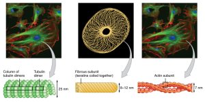
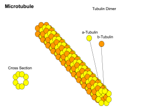 Microtubules, the largest cytoskeletal elements at 25 nm diameter, are made of two closely related proteins, called α-tubulin and β-tubulin. These paired proteins (tubulin dimers) are wrapped into a hollow tube as shown in the diagram and stabilized with microtubule-associated proteins (MAPs).
Microtubules have many uses. They are larger in diameter and more rigid than the other cytoskeletal elements. This makes them useful as:
- “railroad tracks” which are used to move vesicles and other large particles from one end of the cell to the other (this is especially important in neurons, which can be up to 2 meters long);
- structures called mitotic spindles which separate the genetic material during cell division (Unit 6);
- the core of movement organelles like the cilium or the flagellum (next objective); among other uses.
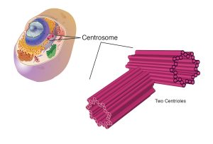
- Microtubules are organized by — wait for it — microtubule-organizing centers (MTOCs). One of the most-studied MTOCs is the centriole, which is surrounded by pericentriolar material. The centriole and its pericentriolar material are together called the centrosome. For example, there is one centrosome at each end of a dividing cell, forming the center of each of the two daughter cells that result from cell division.
Microtubules are organized by — wait for it — microtubule-organizing centers (MTOCs). One of the most-studied MTOCs is the centriole, which is surrounded by pericentriolar material. The centriole and its pericentriolar material are together called the centrosome. For example, there is one centrosome at each end of a dividing cell, forming the center of each of the two daughter cells that result from cell division.
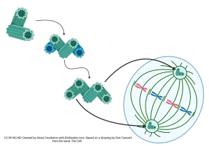
The microtubules growing out of the centrosome are called the mitotic spindle. This spray of microtubules, one from each end of the dividing cell, attaches to the duplicated genetic material packaged into chromosomes and drags the chromosomes to the region that will become each of the daughter cells. We’ll see these mitotic spindles again when we study mitosis in Unit 6.
Intermediate Filaments
Intermediate filaments are, not surprisingly, intermediate in size between microtubules and microfilaments, about 10 nm in diameter. They are made up of a variety of different proteins, including keratin (the major protein of skin cells) and dozens of others.
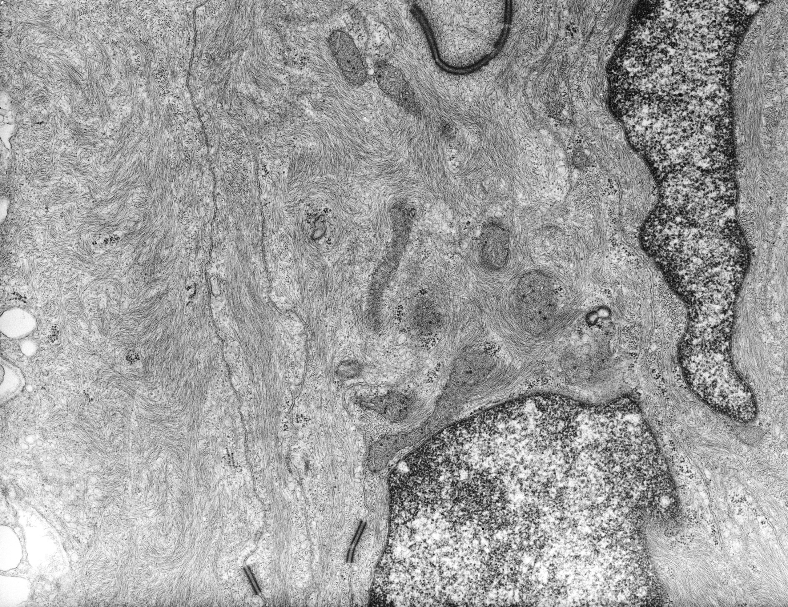 This electron micrograph shows intermediate filament proteins as gray threads helping to hold the shape of a cell that prevents water from entering the frog’s skin.
Another type of nervous system cell is a structural cell, the glial cell. Glial cells contain glial fibrillary acidic protein (GFAP), another of the intermediate filament proteins, again seen as gray threads running through this glial cell visualized by electron microscopy.
Microfilaments (Actin Filaments)
Microfilaments, made up of the protein actin, are the smallest cytoskeletal elements at about 8 nm diameter. They are also the most flexible, in both senses of the word, and are seen in many different contexts. For example, in Unit 10, we will see actin as a major protein that helps muscle cells contract.
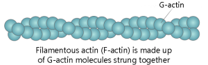
Actin is made up of single protein balls (called globular actin, or G-actin) which can be assembled into fibers, in the style of a strand of pearls. The resulting filamentous actin is called F-actin. Like microtubules, microfilaments are dynamic structures which are constantly being built at one end and broken down at the other as the cell remodels.
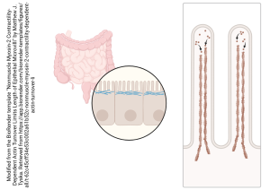
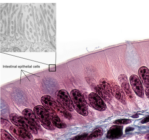
Cells involved in absorption, like the intestinal lining, can increase their surface area by adding numerous finger-like projections called microvilli (sing. microvillus). On the apical (top) surface of the cell, these increase the cell’s surface area, which gives the cell more room for protein pumps, carriers, and enzymes that are needed to aid in absorption along the luminal (open) surface of the gut tube. Each microvillus has a core of actin microfilaments to hold its structure.
Media Attributions
- U05-029 Cytoskeleton © Barnett, Cierra Memphis is licensed under a CC BY-NC-ND (Attribution NonCommercial NoDerivatives) license
- U05-028 fibroblast cytoskeleton © NIH Image Gallery is licensed under a CC BY-NC (Attribution NonCommercial) license
- U05-030 cytoskeleton OpenStax © Betts, J. Gordon; Young, Kelly A.; Wise, James A.; Johnson, Eddie; Poe, Brandon; Kruse, Dean H. Korol, Oksana; Johnson, Jody E.; Womble, Mark & DeSaix, Peter is licensed under a CC BY (Attribution) license
- U05-031 Microtubule_Structure © Lydiawc1020 is licensed under a CC BY-SA (Attribution ShareAlike) license
- U05-032 centrosome © National Human Genome Research Institute is licensed under a Public Domain license
- U05-033 Centriole Replication (version 2) © Crookston, Alexa is licensed under a CC BY-NC-ND (Attribution NonCommercial NoDerivatives) license
- U05-035 intermediate filaments cell image library 9311 © Michaels, JE and Tornheim, PA is licensed under a Public Domain license
- U05-036 actin © Zlir'a adapted by Jordan West is licensed under a CC BY-SA (Attribution ShareAlike) license
- U05-037 actin in microvilli © Tyska, Matthew J. is licensed under a CC BY-NC-ND (Attribution NonCommercial NoDerivatives) license
- U05-038 microvilli 5.9c edited to remove stolen content © Berkshire Community College Bioscience Image Library is licensed under a CC BY-NC-SA (Attribution NonCommercial ShareAlike) license

.jpg)