Structural Classification of Epithelial Tissues
Objective 7.3
7.3.1 Describe unique structural features of the eight epithelial types.
7.3.2 Describe the function and give an example of where each type of epithelial tissue is found.
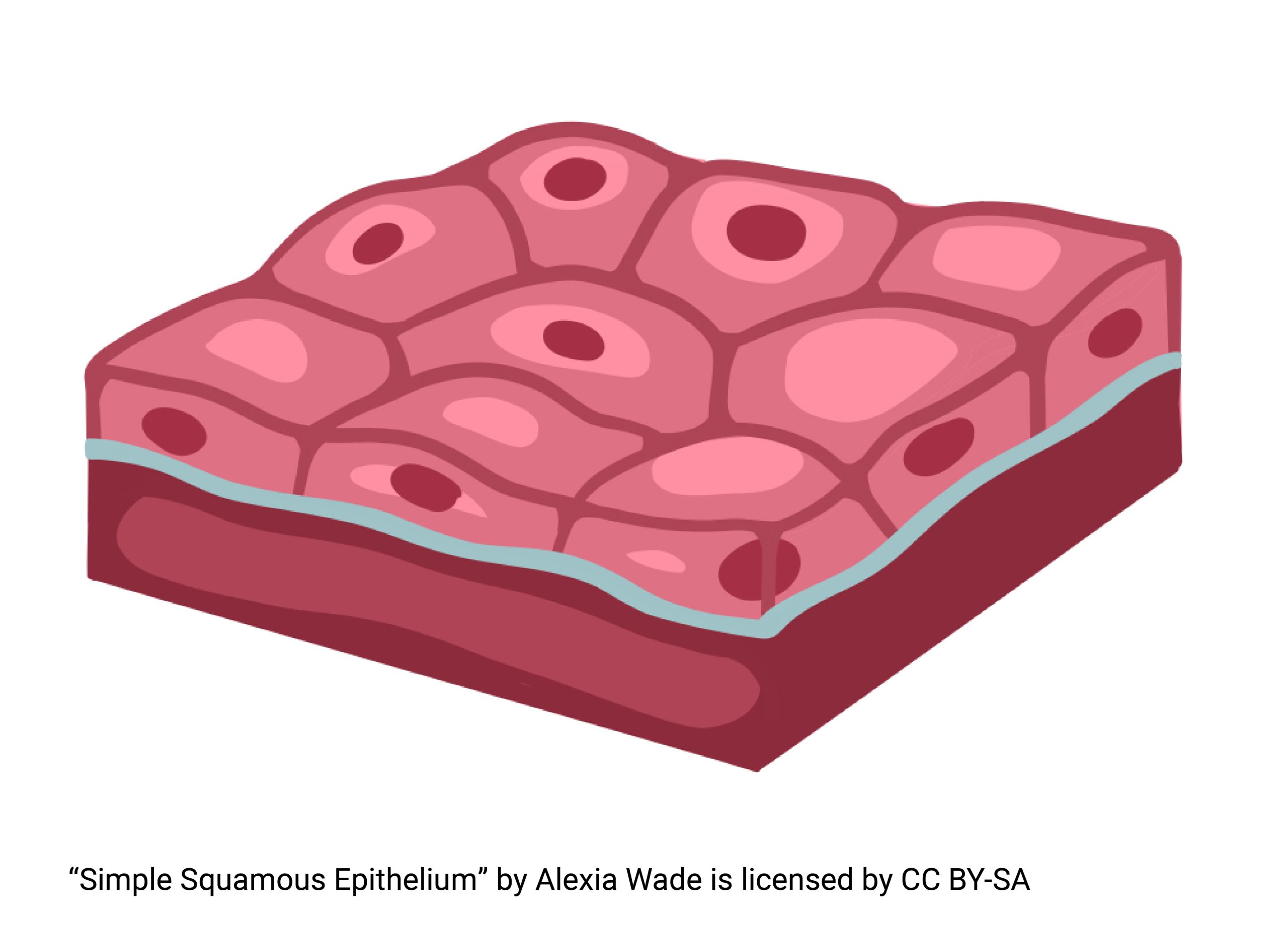
Simple Squamous Epithelium
A simple squamous epithelium consists of a single layer of cells that are thin and flat. It is used where substances need to diffuse across the epithelium, or where filtration is taking place. This type of arrangement limits the obstacles to these passive transport processes. Examples of locations for this type of epithelium include the lining of the heart and blood vessels. Gases such as oxygen and carbon dioxide, and nutrients such as glucose, must be able to freely diffuse across this lining. Trying to get through a brick wall won’t work! We need a single layer of cells for easy diffusion. The air sacs of the lungs (alveoli) are also lined with simple squamous epithelium. In these sacs, oxygen is diffusing from outside air that was just inhaled into tiny blood vessels lining the air sacs. Carbon dioxide gas, a waste product from cellular respiration, must diffuse from the cells of the body to vessels that will carry this back to the lungs (and be exhaled, which plants love)! Again, only a single layer of cells will do for easy diffusion. 
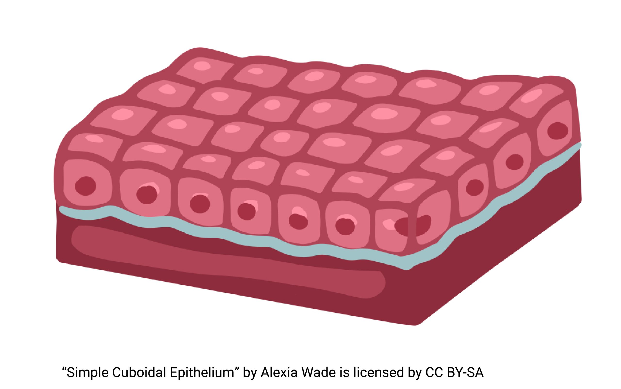
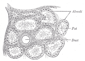
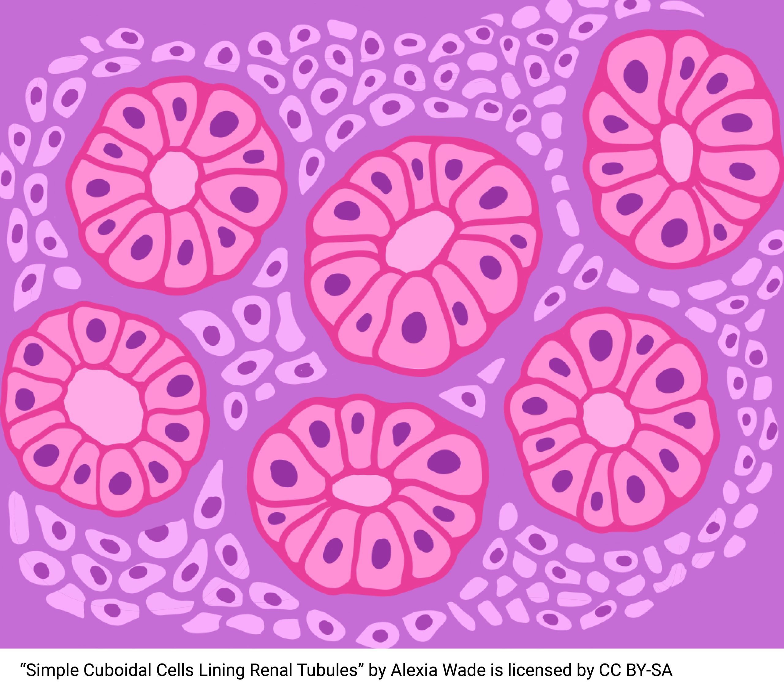
Simple Cuboidal Epithelium
Simple cuboidal epithelium is cube- or square box-shaped. It’s found where secretion or absorption is taking place, as in glandular tissue. We find this tissue in the lining of the kidney tubules. Everything in the blood passes through a three-layer filter in the kidneys. Everything that is small enough gets through (everything but proteins and cells). The problem is the body needs to reclaim much of what passes through, like glucose, for example. The cells need this glucose for energy. Simple cuboidal epithelium is structured to reabsorb this glucose, and other needed substances, back into the bloodstream. The body may want to adjust the blood by getting rid of additional substances in the urine. For example, typically cells in the body generate excess hydrogen ions. Simple cuboidal epithelium can absorb these ions in the kidney tubules, allowing the body to get rid of extra acid. Simple cuboidal epithelium is also found lining the ducts of many exocrine glands; more on these types of glands later. 
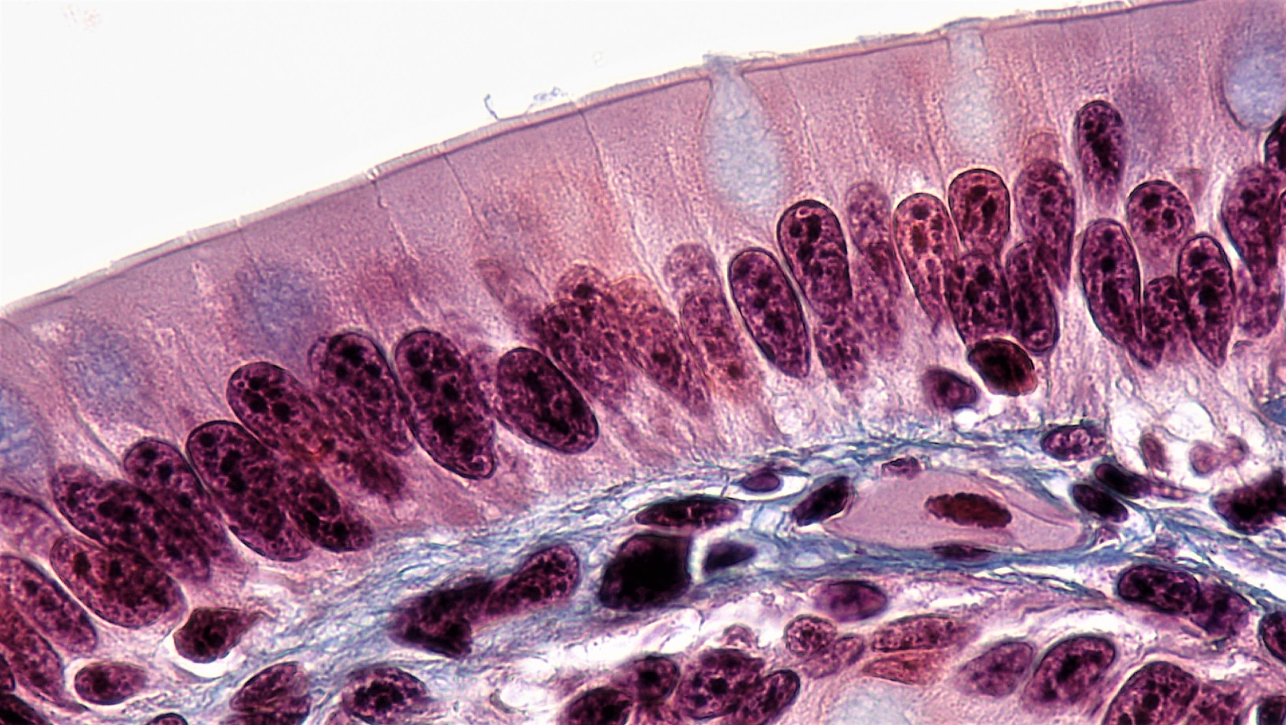
Simple Columnar Epithelium
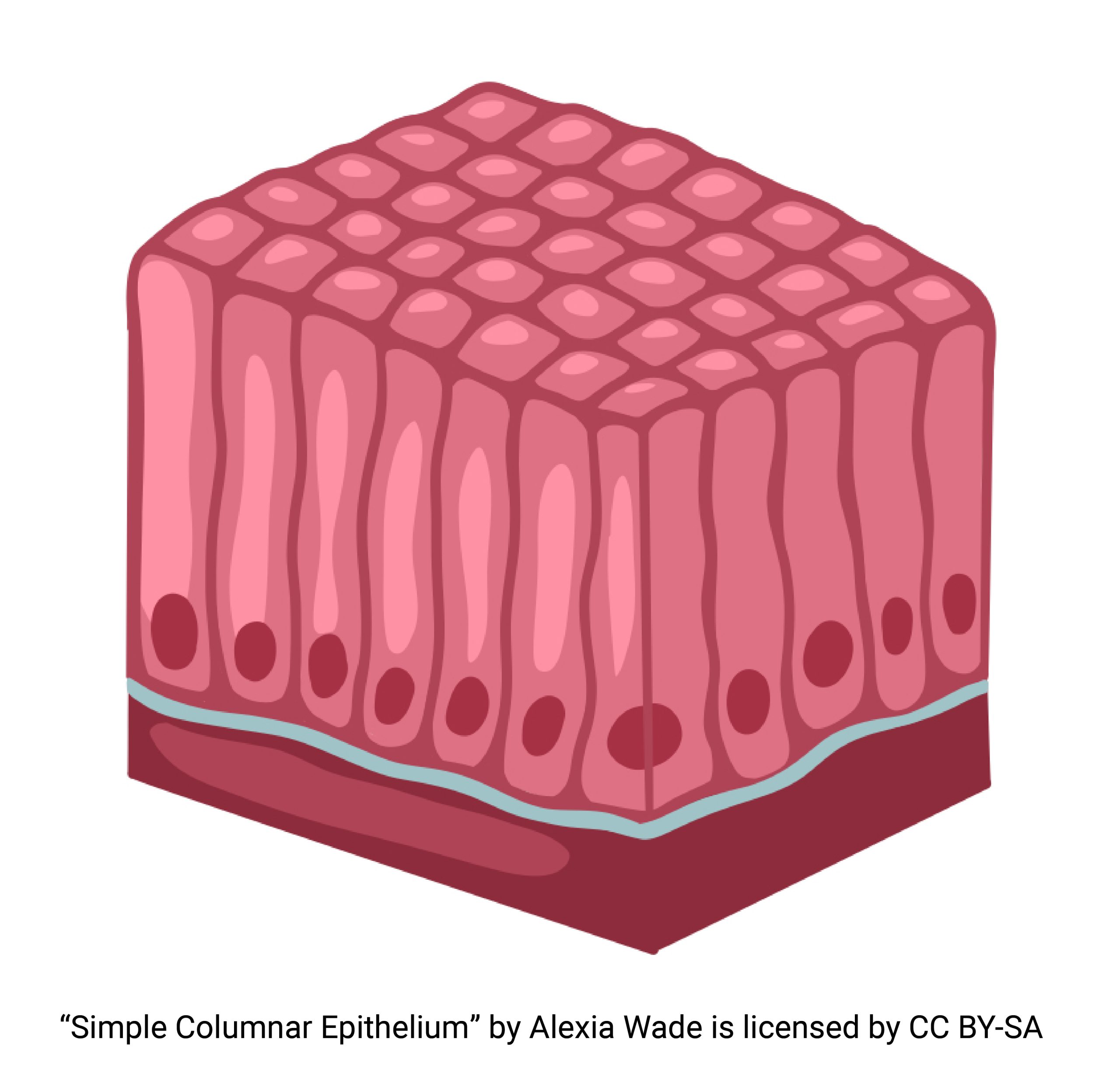
There are two types of simple columnar epithelium. One has cilia on its apical surface. The other does not have cilia. 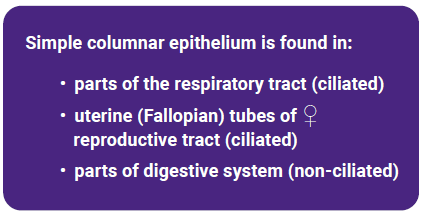
The type with cilia can move mucus and substances trapped in mucus, using the wave-like motion we called the metachronal wave in Unit 5. Together the cilia, the mucus, and the substances trapped in mucus form the mucociliary escalator that lifts material out of the lungs. We find this type anywhere we need to move substances: in the respiratory tract, in the digestive tracts, and in the reproductive tract (uterine tubes) of the female.
The type without cilia generally has microvilli to increase the surface area on the apical surface and is used in the gut. The intestines are lined with a simple columnar epithelium with plentiful microvilli. This is used for breakdown and absorption of nutrients.
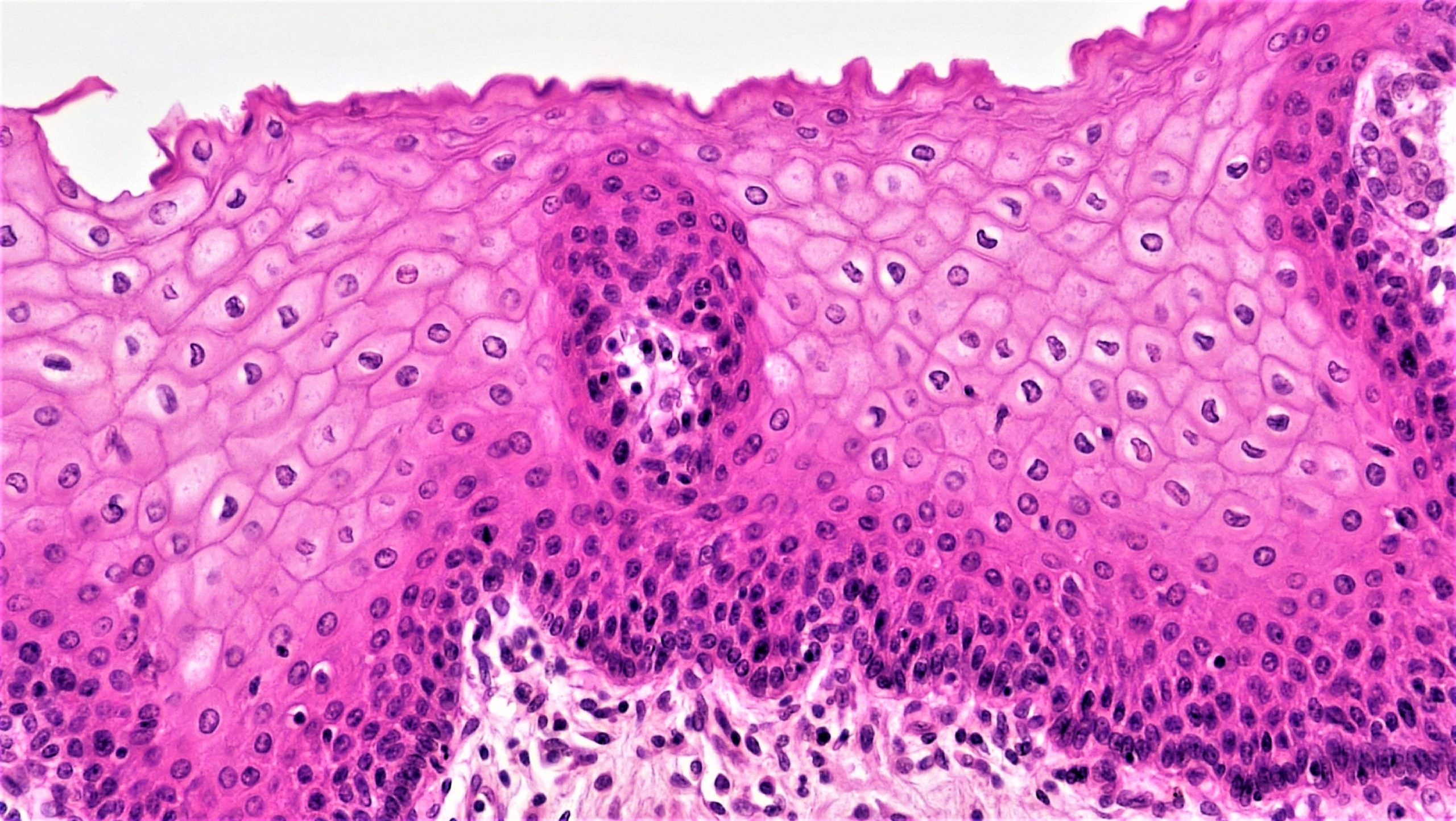
Stratified Squamous Epithelium
Multiple layers of cells, which are thin and flat, are used where we need protection. Almost every organ that meets the outside world needs protection. The skin, the largest organ of the human body, forms a tight barrier to the outside world. The most superficial layer of the skin consists of 25-30 layers of cells arranged in a stratified squamous epithelium. Skin cells produce a fibrous protein, keratin, that helps with cellular adherence and forms a protective layer on the outside of the skin. The esophagus also contains stratified squamous epithelium but this type does not contain keratin. This epithelium helps protect the body from Doritos eaten too rapidly, and any other sharp pointy objects. We‘ll study the skin (integument) in some detail in the next unit as we begin our study of body systems. 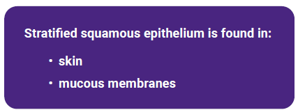
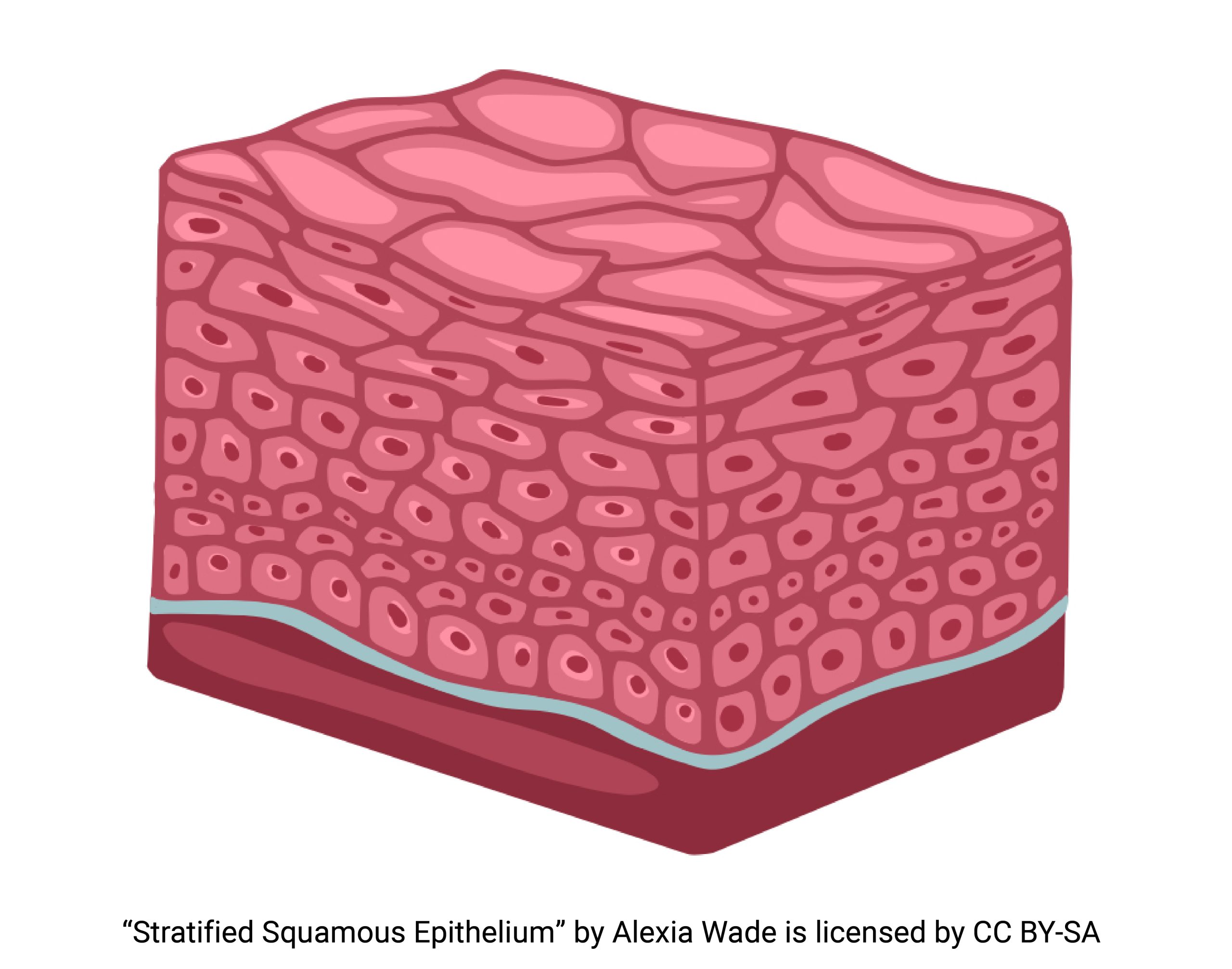
(Notice that it is named after the squamous cells on the apical surface, not the squarish cells at the basal surface.)
Stratified Cuboidal Epithelium
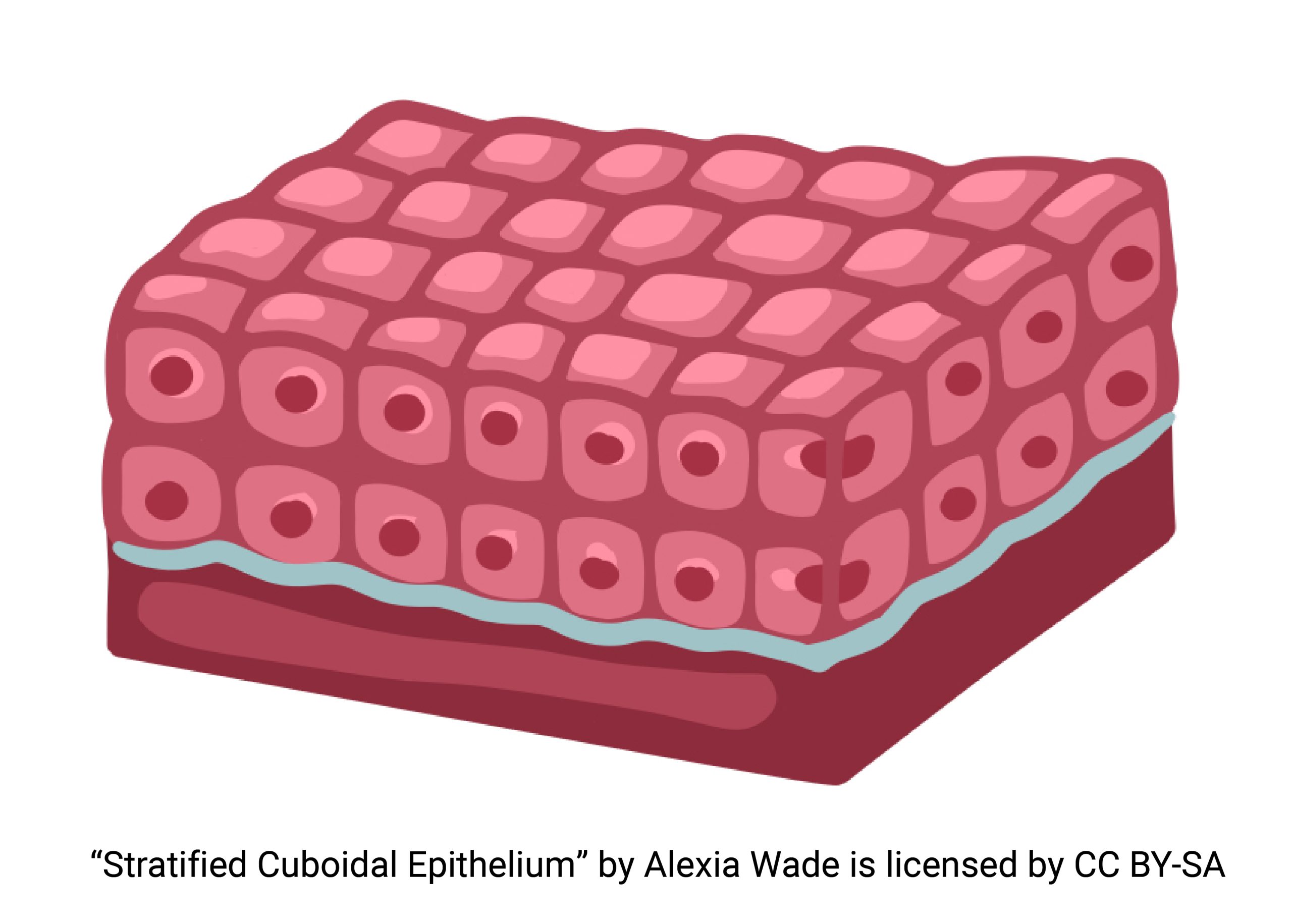
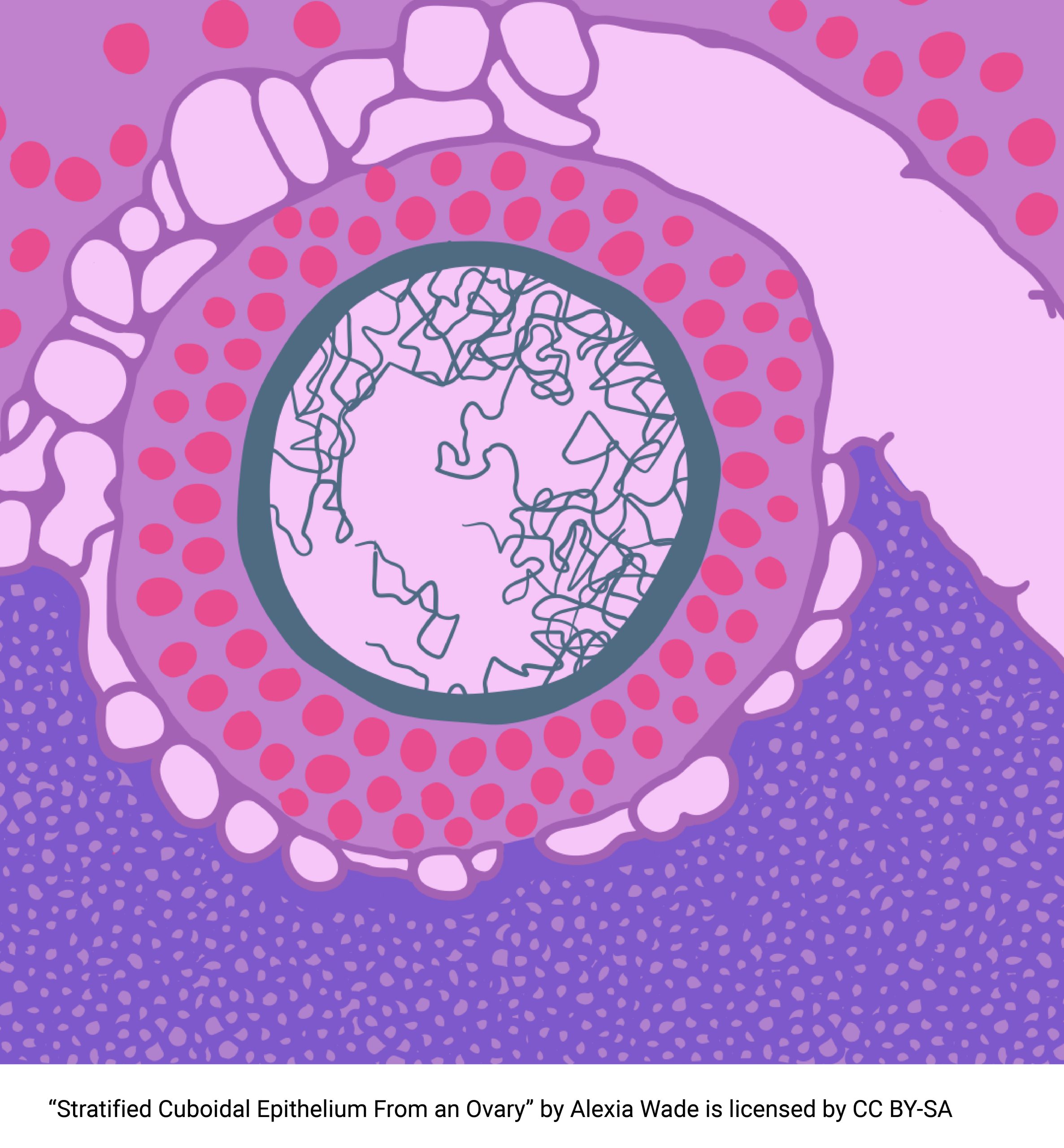
Stratified cuboidal epithelium, with two or more layers of cube-shaped cells, is found in sweat gland ducts, pancreatic ducts and salivary glands. Like a simple cuboidal epithelium, it has a secretory function.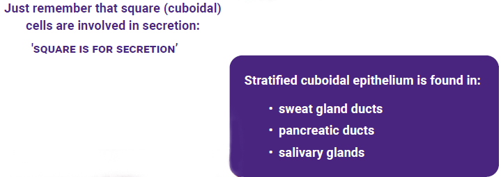
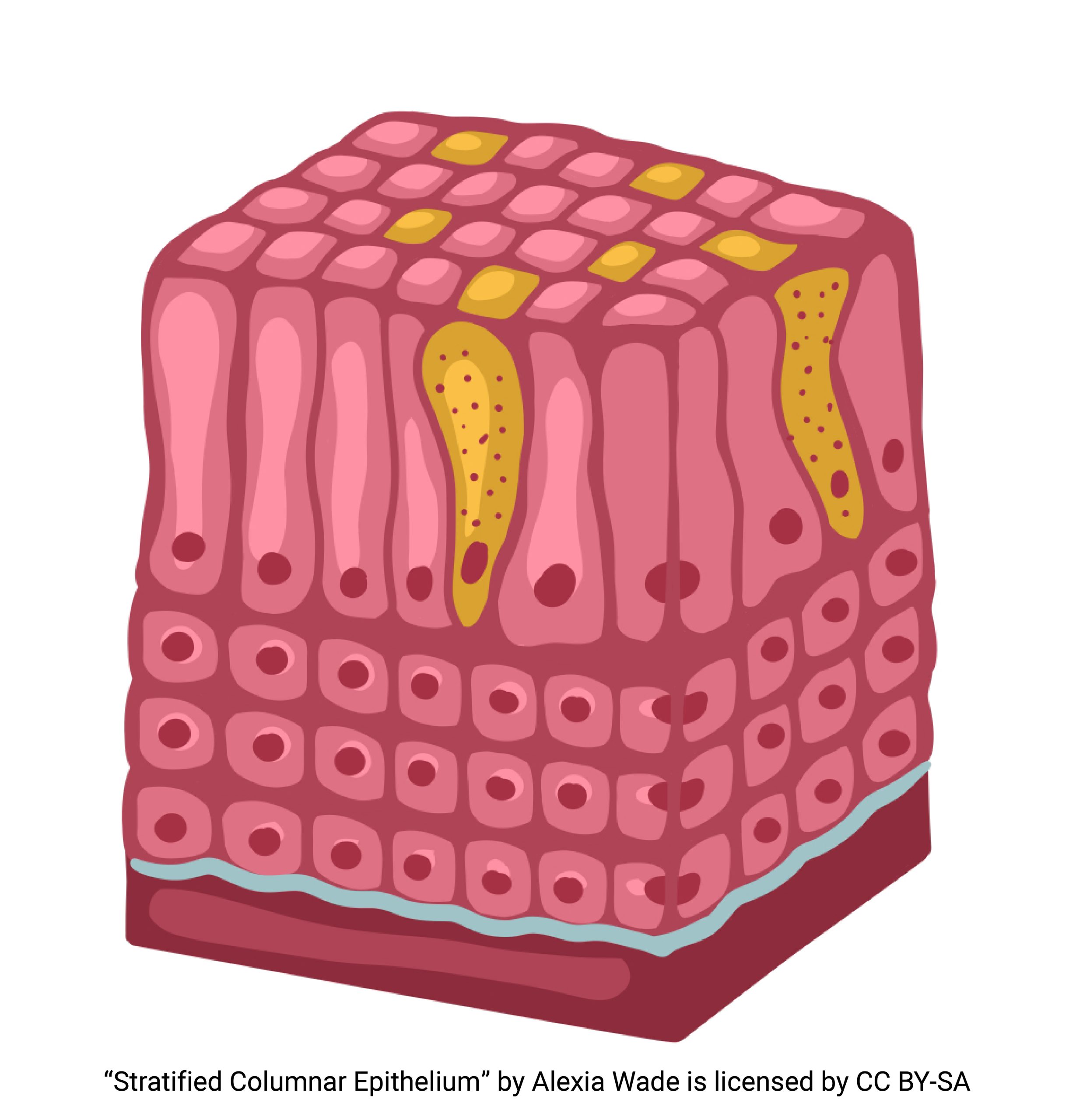
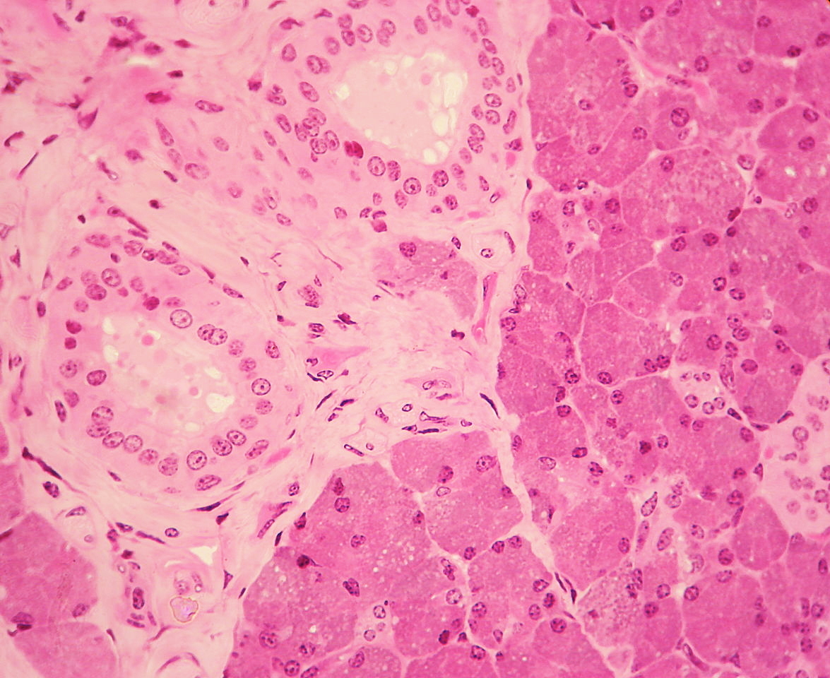
Stratified Columnar Epithelium
Stratified columnar epithelium consists of cells that are taller than they are wide on the apical surface. Although the cells in the basal layers may have a different shape, remember that we name the shape of the cells (columnar) after the cells on the apical surface and these are columnar in shape. Stratified columnar epithelia are not common in the body. Part of the lining of the urethra is composed of stratified columnar epithelium as well as excretory ducts in the esophagus and part of the membrane (conjunctiva) lining the eye. 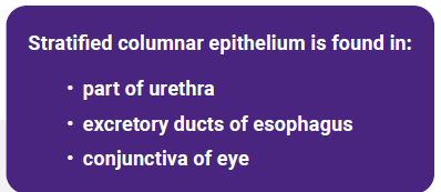
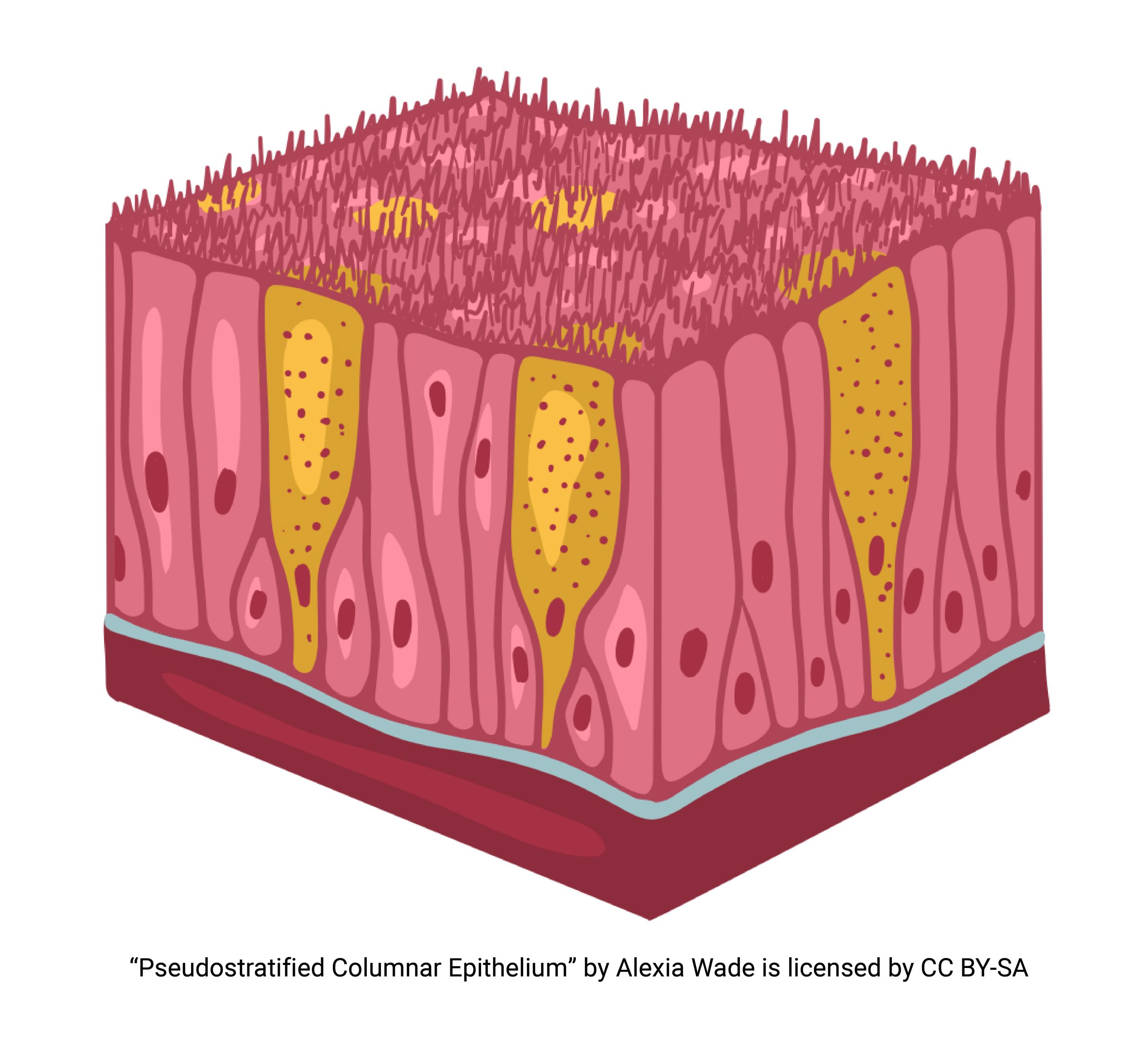
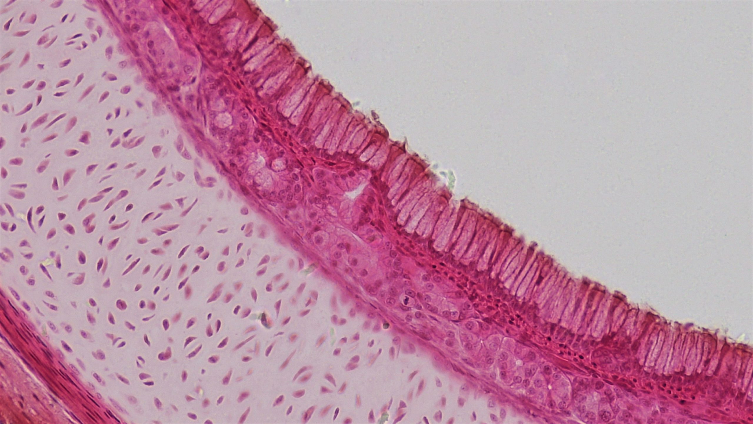
Pseudostratified Columnar Epithelium
Pseudostratified columnar epithelium is unusual. The tissue appears to be stratified but all of the cells are in contact with the basement membrane, but not all extend to the apical surface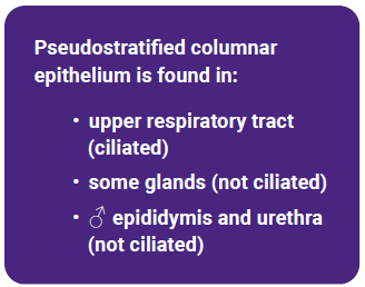
One place it is found, with cilia, is in the upper respiratory tract, where goblet cells secrete mucus to trap dust and invaders, so they can be moved up to the throat by the cilia of the mucociliary escalator. Non-ciliated pseudostratified columnar epithelium is found in some glands, and in the epididymis and urethra of the male where it lines the tubes that conduct sperm and urine.
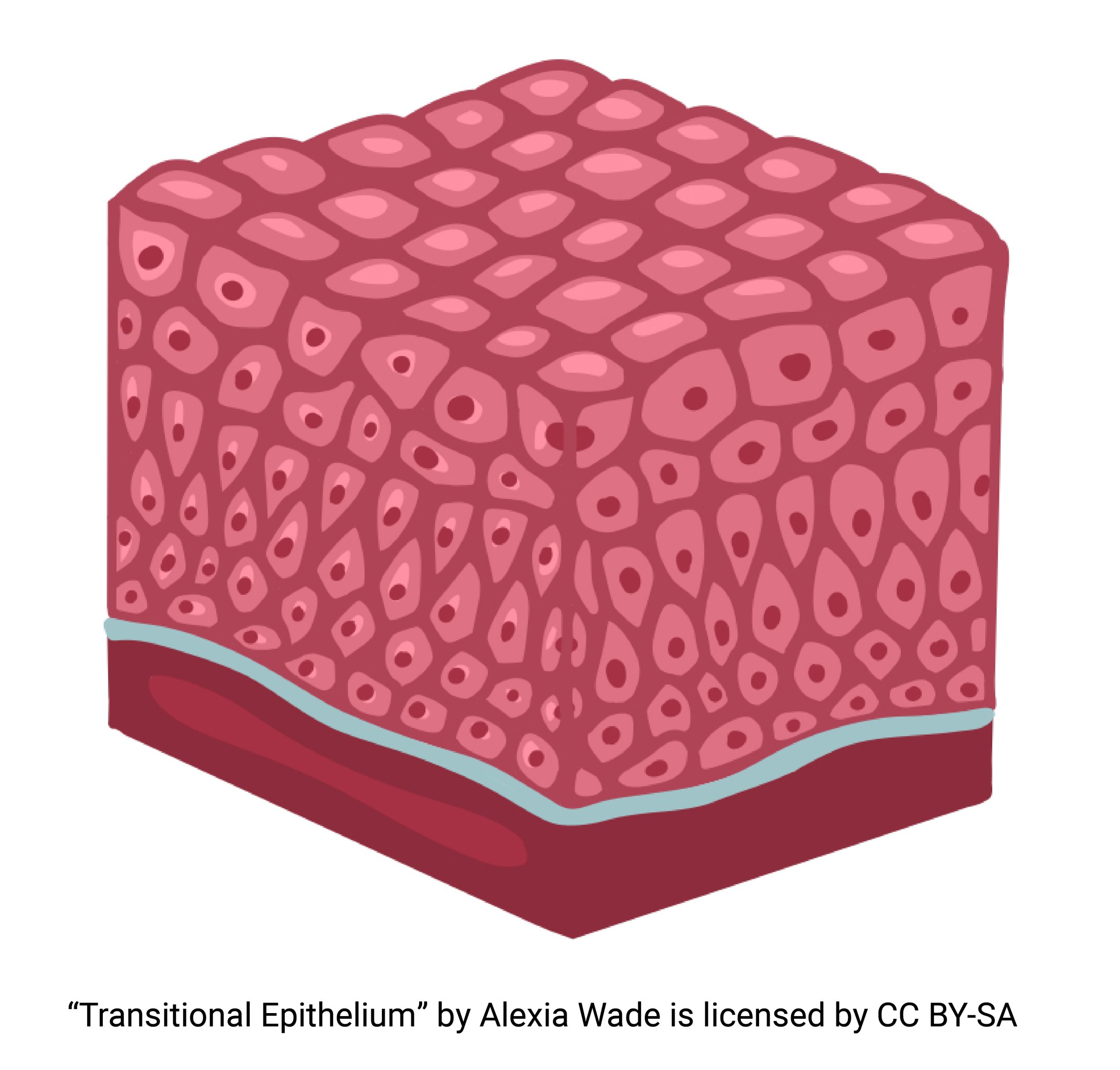
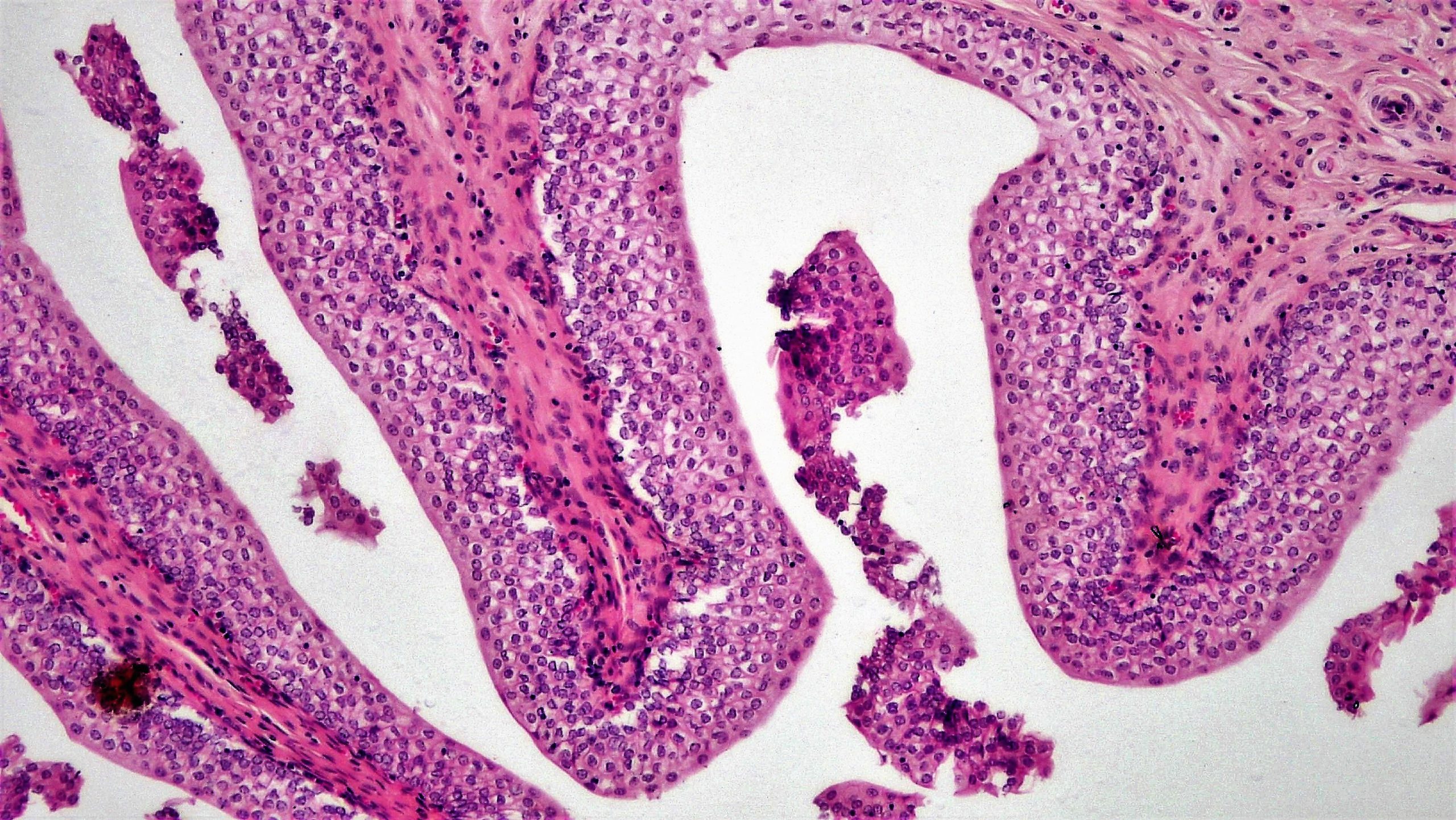
Transitional Epithelium
Transitional epithelium is a term used when the shape of cells, and their layering, changes depending on whether the organ is stretched or contracted. The only examples of such an epithelium are found in the urinary bladder and nearby tubing (ureters and urethra). The cells here take on a squarish shape when the bladder is small and are stretched out flat when the bladder is large.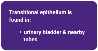
Media Attributions
- U07-008 text box © Bizzell, Lizz is licensed under a CC BY-SA (Attribution ShareAlike) license
- U07-009 © Wade, Alexia is licensed under a CC BY-SA (Attribution ShareAlike) license
- 23_13 © OpenStax is licensed under a CC BY-SA (Attribution ShareAlike) license
- U07-013 text box © Bizzell, Lizz is licensed under a CC BY-SA (Attribution ShareAlike) license
- U07-011 © Wade, Alexia is licensed under a CC BY-SA (Attribution ShareAlike) license
- U07-012 Gray1173 © Carter, Henry Vandyke is licensed under a Public Domain license
- U07-014 © Wade, Alexia is licensed under a CC BY-SA (Attribution ShareAlike) license
- U07-015 © Wade, Alexia is licensed under a CC BY-SA (Attribution ShareAlike) license
- U07-017 text box © Bizzell, Lizz is licensed under a CC BY-SA (Attribution ShareAlike) license
- U07-018 simple columnar 27854453998_2616880b28_o © Berkshire Community College Bioscience Image Library is licensed under a Public Domain license
- U07-019 text box © Bizzell, Lizz is licensed under a CC BY-SA (Attribution ShareAlike) license
- U07-020 © Wade, Alexia is licensed under a CC BY-SA (Attribution ShareAlike) license
- U07-021 stratified squamous epithelium 40230842160_a605a7594b_o © Berkshire Community College Bioscience Image Library is licensed under a Public Domain license
- U07-023 text boxes © Bizzell, Lizz is licensed under a CC BY-SA (Attribution ShareAlike) license
- U07-022 © Wade, Alexia is licensed under a CC BY-SA (Attribution ShareAlike) license
- U07-024 © Wade, Alexia is licensed under a CC BY-SA (Attribution ShareAlike) license
- U07-026 text box © Bizzell, Lizz is licensed under a CC BY-SA (Attribution ShareAlike) license
- U07-025 © Wade, Alexia is licensed under a CC BY-SA (Attribution ShareAlike) license
- U07-027 WVSOM_Parotid_Gland1 © Wbensmith is licensed under a CC BY (Attribution) license
- U07-029 text box © Bizzell, Lizz is licensed under a CC BY-SA (Attribution ShareAlike) license
- U07-028 © Wade, Alexia is licensed under a CC BY-SA (Attribution ShareAlike) license
- U07-030 26915091897_b28bcbae1c_o © Berkshire Community College Bioscience Image Library is licensed under a Public Domain license
- U07-033 text box © Bizzell, Lizz is licensed under a CC BY-SA (Attribution ShareAlike) license
- U07-032 © Wade, Alexia is licensed under a CC BY-SA (Attribution ShareAlike) license
- U07-034 41850591822_7a2914b3ae_o © Berkshire Community College Bioscience Image Library is licensed under a Public Domain license
