Sliding Filament Theory
Objective 10.4
10.4.1 Describe the organization of a sarcomere.
10.4.2 Diagram the location of myosin and actin filaments.
10.4.3 Explain what is meant by “sliding filament model.”
We have already discussed the way in which thousands of sarcomeres shorten to allow myofibrils to shorten, which collectively shortens the muscle cells, which shortens fascicles, which shorten muscles, giving us contraction. But how do the sarcomeres actually shorten? This objective will explain the way in which individual sarcomeres are able to shorten and return to their original shape.
We saw in light micrographs of skeletal muscle that the striations that give striated muscle its name were sarcomeres, the contractile unit of muscle. The alternating areas of light and dark come about because of the arrangement of actin and myosin within each sarcomere.
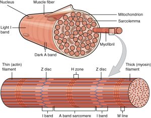
The actin filaments are held together at the Z disc (line) by protein called actinin. The Z-disc is the dark line that splits the light areas in the micrographs seen here. The Z disc defines the borders of the sarcomere. Using electron microscopy, we can see the relationship between the thick filaments of myosin and the thin filaments of actin. There are regions where only actin filaments are found near the Z-discs, regions where only myosin filaments are found near the middle of the sarcomere or the M-line, and regions where they overlap. These regions changes as the sarcomeres shorten or spring back to their original shape after contraction.
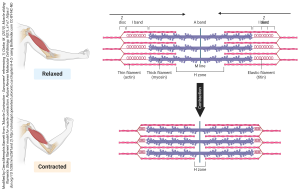
We will see in more detail later how the centrally located myosin proteins reach across the gap and bind to the actin proteins and pull them towards the center of the sarcomere. As more myosin heads bind and pull in on actin, the sarcomere continues to decrease in length. After contraction, the actin binding sites on actin are covered and structural proteins like titin and elastin help return the sarcomere to its original shape. The process is repeated every time a muscle contracts. This is referred to as the sliding filament model.
It is important to note the extent of overlap between the actin filaments and the myosin filaments. The amount of overlap has a direct impact on the strength, or tension that can be generated by the contracting sarcomere. This relationship is known as the length-tension relationship.
Let’s start in the middle of the graph, where the most force can be generated. Notice the green lines representing the actin thin filaments, and the brown hairy thing representing the myosin thick filaments. The “hairs” are the myosin heads. In the middle, at a sarcomere length of about 2.5 μm, there are lots of opportunities for myosin to interact with actin (lots of potential points of contact between the brown “hairs” and the green lines).

Now look to the right side of the graph. If the sarcomere is stretched out to about 4 μm, then there is no overlap between the thin and thick filaments and no opportunity for myosin to generate force.
The hardest (and probably least important) to grasp is the left side of the graph. Here, the actin thin filaments overlap with each other; this also reduces the ability of myosin and actin to interact and generate force.
Muscle force, as we will see in Objective 6, is dependent on the interaction between actin and myosin molecules.
Media Attributions
- 1002_Organization_of_Muscle_Fiber © Betts, J. Gordon; Young, Kelly A.; Wise, James A.; Johnson, Eddie; Poe, Brandon; Kruse, Dean H. Korol, Oksana; Johnson, Jody E.; Womble, Mark & DeSaix, Peter is licensed under a CC BY (Attribution) license
- U10-021 Sliding Filament Model © Barnett, Cierra Memphis is licensed under a CC BY-NC-ND (Attribution NonCommercial NoDerivatives) license
- U10-022 1011_Muscle_Length_and_Tension © Betts, J. Gordon; Young, Kelly A.; Wise, James A.; Johnson, Eddie; Poe, Brandon; Kruse, Dean H. Korol, Oksana; Johnson, Jody E.; Womble, Mark & DeSaix, Peter is licensed under a CC BY (Attribution) license
