Skin Pigment
Objective 8.3
8.3.1 Identify and describe the three main skin pigments.
8.3.2 Know which layer of the skin contains each pigment.
The color of the skin comes from three classes of pigments: melanin, hemoglobin, and carotene.
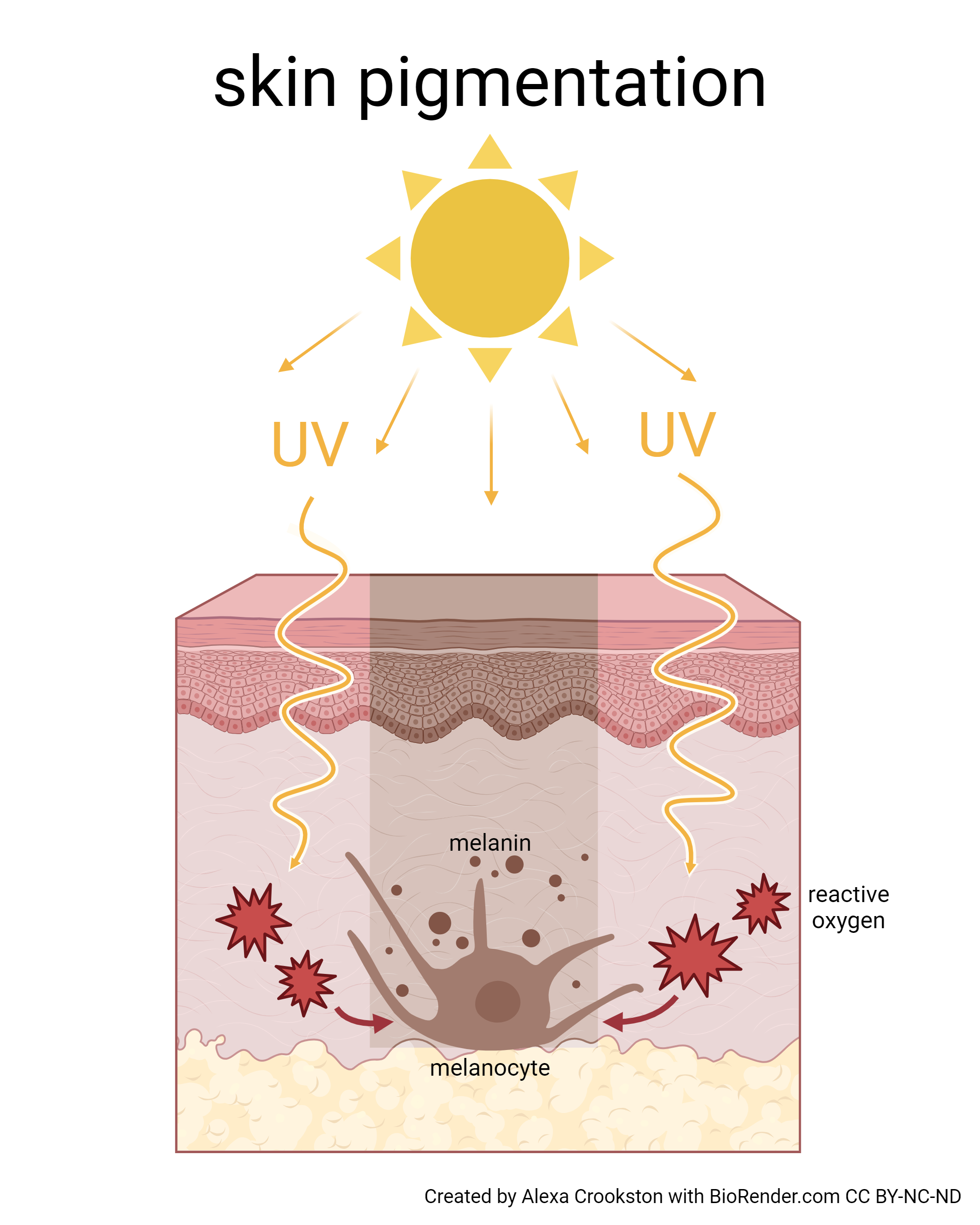
Melanin: Recall that melanin is produced by cells called melanocytes, which are found scattered throughout the stratum basale of the epidermis.
There are two kinds of melanin (a pigment that can absorb UV light):
- Pheomelanin, which is yellow to red; (freckles)
- Eumelanin, which is brown to black.
In hair, pheomelanin and eumelanin granules are scattered among the keratin fibers. In skin, these pigments are produced in cells called melanocytes. Almost everyone has roughly the same number of melanocytes; where people differ in skin color is in the amount of pigment in each melanocyte. Melanin is made from the amino acid tyrosine in an organelle called the melanosome. The pigment is transferred to keratinocytes by melanosomes giving hair and epidermis their color.
Exposure to the UV rays of the sun or a tanning salon causes melanin to be manufactured and built up in keratinocytes, as sun exposure stimulates keratinocytes to secrete chemicals that stimulate melanocytes. The accumulation of melanin in keratinocytes results in the darkening of the skin, or a tan. This increased melanin accumulation protects the DNA of epidermal cells from UV ray damage and the breakdown of folic acid, a nutrient necessary for our health and well-being. In contrast, too much melanin can interfere with the production of vitamin D, an important nutrient involved in calcium absorption. Thus, the amount of melanin present in our skin is dependent on a balance between available sunlight and folic acid destruction, and protection from UV radiation and vitamin D production.
It requires about 10 days after initial sun exposure for melanin synthesis to peak, which is why pale-skinned individuals tend to suffer sunburns of the epidermis initially. Dark-skinned individuals can also get sunburns, but are more protected than are pale-skinned individuals. Melanosomes are temporary structures that are eventually destroyed by fusion with lysosomes; this fact, along with melanin-filled keratinocytes in the stratum corneum sloughing off, makes tanning impermanent.
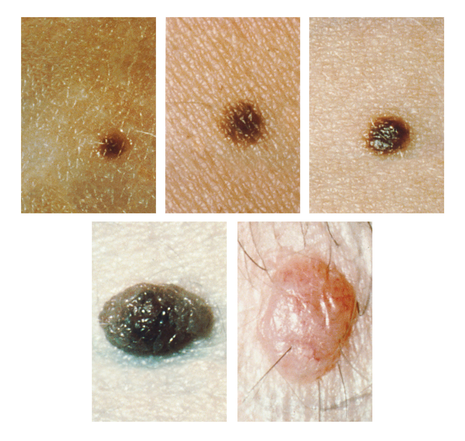
Too much sun exposure can eventually lead to wrinkling due to the destruction of the cellular structure of the skin, and in severe cases, can cause sufficient DNA damage to result in skin cancer. When there is an irregular accumulation of melanocytes in the skin, freckles appear. A mole, or nevus, is a larger mass of melanocytes. Although most are benign, they should be monitored for changes that might indicate the presence of cancer (see figure at the right and Objective 11).
Melanocytes are normally scattered more-or-less evenly throughout the epidermis, mostly in the deeper layers (spinosum and basale). In light-skinned persons, little melanin is made and stored in melanocytes. In these people, the color of the skin is predominantly due to hemoglobin.
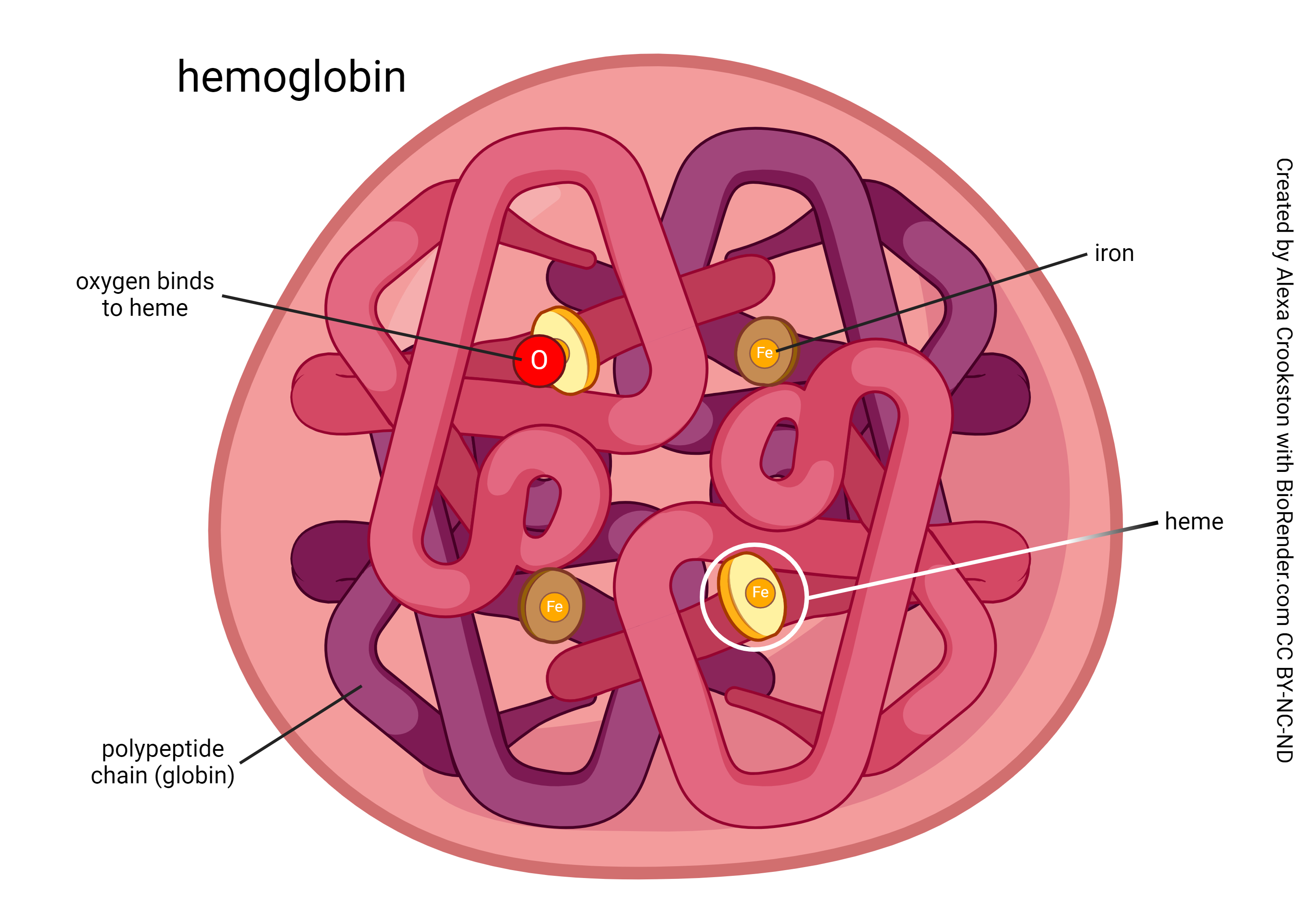
Hemoglobin: a red pigment in the blood that gives the skin of light-skinned individuals a pinkish cast as it circulates through capillaries in the dermis. The more blood is present near the surface of these individuals, the pinker the skin becomes (i.e. blushing or flushing).
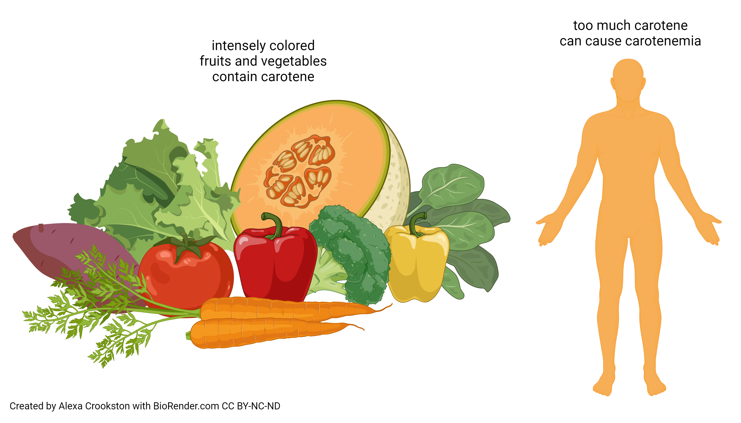
Carotene: Intensely colored fruits and vegetables contain a fat-soluble pigment called carotene. Carotene is a precursor of Vitamin A and can deposit in the skin. Excessive consumption of these fruits and vegetables can cause carotenemia, carotene in the blood causing the skin to appear orange.
In some diseases, such as liver failure, the pigment bilirubin accumulates in the skin, giving it a yellow color. This is not a normal skin pigment; the symptom of yellow skin (particularly easy to see in the “whites” or sclera of the eye) is called jaundice (French, jaune, “yellow”).
Media Attributions
- U08-008 Skin Pigmentation © Crookston, Alexa is licensed under a CC BY-NC-ND (Attribution NonCommercial NoDerivatives) license
- U008-009 Moles © National Cancer Institute is licensed under a Public Domain license
- U08-010 Hemoglobin © Crookston, Alexa is licensed under a CC BY-NC-ND (Attribution NonCommercial NoDerivatives) license
- U08-011 Carotene, Carotenemia © Crookston, Alexa is licensed under a CC BY-NC-ND (Attribution NonCommercial NoDerivatives) license
