Muscle Organization and Structure
Objective 10.3
10.3.1 Describe the levels of skeletal muscle organization, from individual cells to muscles to muscle groups.
10.3.2 Identify the connective tissue layers around cells, fascicles and muscles and describe the way in which a muscle attaches to a bone or other muscle.
10.3.3 Explain the role of T tubules and sarcoplasmic reticulum within the cell.
The smallest unit of skeletal muscle is the sarcomere, a regular arrangement of actin and myosin filaments plus structural proteins to hold things in place.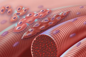
Many thousands of sarcomeres are lined up in a myofibril, a long strand of sarcomeres arranged end-to-end.
Several dozen myofibrils are found inside of each muscle cell, or muscle fiber. This is called a syncytium and is the result of embryonic muscle cells fusing to form a long tube.
The cell membrane of the skeletal muscle cell has the rather grand and strange name sarcolemma. It’s just like the cell membrane of any other cell; no one knows why this strange name persists.
Two Latin names appear over and over again in this Unit.
Sark or sarx (Greek σάρξ) means “flesh” in Greek, and so all the “sarco–” words come from that root: sarcoplasm, sarcolemma, sarcomere.
μῦς orMus, which became myo–, is the Greek word for mouse! The action of muscles like the biceps was thought to look like a mouse, probably by the same Greeks who named the constellations. They had a very active imagination.
Similarly, instead of the cytoplasm of muscle cells, we refer to the sarcoplasm.
muscle fiber = muscle cell
sarcolemma = cell membrane = plasmalemma
sarcoplasm = cytoplasm
sarcoplasmic reticulum = smooth endoplasmic reticulum
Lower motor neurons run throughout the muscle, but do not extend deep into each individual muscle cell. An elaborate system of tubules functions to relay the signal from the neuron resting just above the cell membrane to all of the sarcomeres inside the cell. These tubules, called T tubules, are like little folds of plasma membrane that dip into the cell. Flanking the T tubules on each side are tiny bags of calcium known as sarcoplasmic reticula. The sarcoplasmic reticulum is a specialized form of smooth endoplasmic reticulum which stores calcium ions (Ca2+) and releases them when needed. They are necessary in the contraction process.
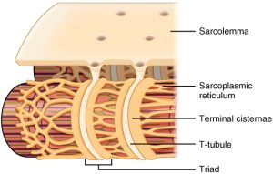
Because the electrical and calcium management systems of the muscle cell must work together, as we’ll see later, they are in close proximity to each other. One cylindrical T tubule is flanked by two flattened sacs of smooth endoplasmic reticulum in a standardized arrangement called a triad.
In order to function properly, the contractile machinery inside muscle cells must be connected to tendon and bone. This connection is made by several levels of connective tissue. Each of these connective tissue layers ends in –mysium, which means “muscle”.
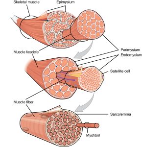
- Endomysium is a thin sheet of connective tissue that surrounds each muscle cell, lying just over the muscle cell, lying just over the muscle cell membrane (sarcolemma). This innermost connective tissue sheet has the prefix endo– which means “inner”.
- Perimysium is a connective tissue sheet which wraps about a dozen individual muscle cells. These groups of muscle cells, covered by perimysium, are called fascicles and are the smallest visible unit of muscle, seen as the “grain” in fresh meat. The prefix “peri–” means “surrounding”.
- Epimysium is a strong, thick sheet of connective tissue that covers an entire muscle. This is the sheet of dense irregular connective tissue that transitions into the dense regular connective tissue of the tendon. The prefix “epi–” means “on top of”, as in, it is a layer that is on top of the entire muscle.
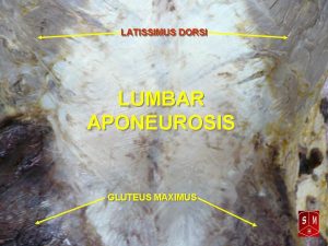
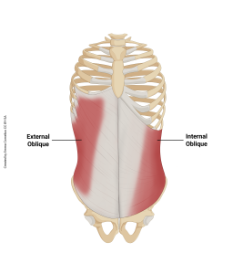
Sometimes the connective tissues of muscle form broad, flat sheets that allow for muscles to attach across wide bone surfaces or to other muscles. These structures are called aponeuroses. Aponeuroses in the body can be found throughout the body, for example; where muscles attach to the skull, where the diaphragm attaches to the central tendon, and anywhere where larger muscles like the abdominal or trapezius muscles make connections. Tendons and aponeuroses have few blood vessels and nerves and therefore appear glossy and whitish in color.
Media Attributions
- myoblast fusion6_21test lines6 © Leja, Darryl NHGRI is licensed under a Public Domain license
- U10-015 1023_T-tubule © Betts, J. Gordon; Young, Kelly A.; Wise, James A.; Johnson, Eddie; Poe, Brandon; Kruse, Dean H. Korol, Oksana; Johnson, Jody E.; Womble, Mark & DeSaix, Peter
- U10-O16a © Betts, J. Gordon; Young, Kelly A.; Wise, James A.; Johnson, Eddie; Poe, Brandon; Kruse, Dean H. Korol, Oksana; Johnson, Jody E.; Womble, Mark & DeSaix, Peter is licensed under a CC BY (Attribution) license
- U10-018 Lumbar_aponeurosis © Anatomist90 is licensed under a CC BY-SA (Attribution ShareAlike) license
- U10-017 aponeurosis and obliques © Cornelius, Emma is licensed under a CC BY-SA (Attribution ShareAlike) license
