Muscle Metabolism
Objective 10.7
10.7.1 Describe the ways in which ATP molecules are used in muscle fibers.
10.7.2 Explain the role of oxygen in ATP production and describe what happens when oxygen levels are low.
10.7.3 Explain the role of other molecules like creatine and myoglobin in energy metabolism.
10.7.4 Differentiate between fast and slow twitch muscle fibers.
With so much discussion of muscle contraction and force, it would be unwise to not explore how the body generates the energy needed to keep our muscles working. This objective will build off our earlier discussion of energy production and cellular metabolism (Unit 5, Objective 2-9). You will also build off of your understanding of the unique characteristics of skeletal, smooth, and cardiac muscle (Unit 7, Unit 10). Now is probably a good time to review those objectives before moving on.
ATP is well understood to be the body’s energy currency. No other system of the body compares to the muscular system in terms of sheer energy expenditure, and for good reason. If skeletal, cardiac or smooth muscles failed to do their jobs we wouldn’t last very long. In order to maintain appropriate levels of energy for muscle use, the body works hard to maintain adequate glycogen deposits within the muscles. About 1500 food calories (1500 Cal) of glycogen are storied in the muscles, with another 500 Cal stored in the liver. It is possible to burn through all of one’s stored glucose. Endurance athletes often experience this and describe it as “hitting a wall.”
The mechanisms of ATP production outside of glucose metabolism are less desirable for quick energy and leave the body to deal with metabolic byproducts that can be harder to manage. Fatty acids can be used as an energy source, but this produces acid and ketones. Proteins can even be used for energy, but the breakdown of proteins, including the muscle proteins actin and myosin, for the purpose of keeping muscles working is a last resort and atypical for a healthy individual.
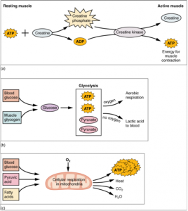
Recall that glycogen is a branched polysaccharide comprised of many individual glucose molecules. These glucose molecules can be broken off and used to produce ATP. When oxygen is present, the total yield from one molecule of glucose is in the ballpark of 36-38 molecules of ATP. Without oxygen, the total yield drops to only two ATP. For that reason, oxygen transport throughout the muscle is extremely important.
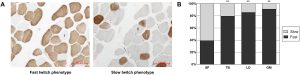
Where only a few short twitches are needed, like in the extraocular muscles of the eye or smaller muscles of the fingers, there is little need for extensive amounts of glycogen or oxygen, therefore myoglobin levels are lower. This contributes to the lighter color of the muscle tissues upon gross examination. These fibers may be referred to as fast twitch, glycolytic fibers because they have a lower demand for ATP and can perform their smaller, quicker movements without relying on aerobic respiration to generate the energy. Conversely, muscles that remain contracted for long periods of time, like those of the back and legs, will have greater amounts of myoglobin and appear darker. These muscle fibers are referred to as slow twitch, oxidative fibers because they contract more slowly and for longer periods of time and primarily rely on aerobic oxidation of glucose for ATP synthesis. A “compromise” type, called fast oxidative-glycolytic (FOG) fibers, is also present.
We can stain for mitochondrial activity to visualize these different fiber types, as shown in the image. The more mitochondrial activity, the darker the staining.
The muscle fibers of the human body combine these three metabolic strategies to varying degrees. All muscle is a mixture of these three types, but one type of fiber can predominate in some muscles that need a lot of that fiber type. Athletic training might also result in some switching of fiber types. Athletes care about fiber twitch type because different sports require different combinations of fiber types. Think of weight lifters compared to table tennis players – they’re going to have very different combinations of fiber types. Groups of muscles can have a varying number of each fiber type, with different fiber types being innervated as part of different motor units to provide the desired type of muscle contraction for a given activity.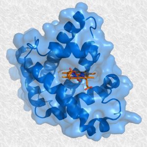
Hemoglobin is an iron containing, oxygen carrying protein found in red blood cells. Muscles make use of a similar molecule, myoglobin, to transport and even store oxygen throughout the muscle. As oxygen need increases in muscle tissue, the myoglobin concentration will increase. Increased amount of myoglobin is why some muscle tissue appears darker than others. The amount of stored myoglobin is a reflection of the type of work the muscle tissue performs.
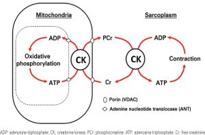
Myoglobin gives muscle tissue some added oxygen accessibility. Another molecule, creatine, has a similar role, only it serves to increase the availability of ATP in muscle tissue. You can think of creatine as a storage container for phosphates. When the muscle tissue is at rest, with ATP excess, the enzyme creatine kinase (CK) combines free phosphates with creatine to create creatine phosphate. When ATP levels are low, creatine phosphate can donate the stored phosphates to ADP molecules, converting them into ATP. Creatine derived energy is a good backup to the stored ATP found in muscle cells.
Like many other cells of the body, muscle cells do not function well in the absence of ATP. For reasons not fully understood, muscles can experience a phenomenon known as fatigue. Current theories explain muscle fatigue as a defensive mechanism to prevent excessive damage and death to muscle fibers. Although the exact biochemical mechanisms of what is happening are not easily explained we know there are at least two sources of muscle fatigue. The first source of fatigue is depletion of ATP due to a lack of carbohydrates, creatine, or the oxygen needed to fuel oxidative metabolism. As muscles attempt to function with depleted ATP and oxygen a number of byproducts can collect in the muscles. These include ADP, lactic acid, and potassium.
The other source of fatigue is the central nervous system. When the master motor neurons of the CNS decide to quit in attempt to avoid damage, the motor system shuts down. This is more likely to happen if (say) you are trying to run a marathon than if you are being chased by a tiger.
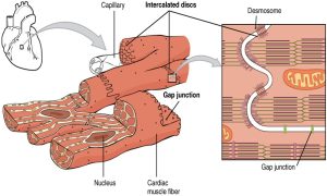
Obviously, cardiac muscle must be able to resist fatigue. Beating over 100,000 times each day with little rest is a perfect recipe for fatigue. Cardiac muscle has high amounts of creatine, myoglobin and mitochondria as well as a dedicated source of oxygenated blood to keep the muscle working around the clock. Without oxygenated blood, the heart muscle cannot work. Only a small fraction of the energy used by cardiac muscle comes from anaerobic metabolism of glucose. The heart is “tuned” to only use oxygen and glucose in oxidative metabolism. If the heart is starved of oxygen, even briefly, it fails. The first signal of this is angina, a pain in the chest; if the condition lasts too long, the heart stops.
Media Attributions
- U10-028 Statue_of_the_Tired_Man © User:Burrows is licensed under a Public Domain license
- U10-029 muscle-metabolism-768×860 © CFCF is licensed under a CC BY-SA (Attribution ShareAlike) license
- U10-030 fiber types © Bao, Tugeqin; Han, Haige; Li, Bei; Zhao, Yiping; Bou, Gerelchimeg; Zhang, Xinzhuang; Du, Ming; Zhao, Ruoyang; Mongke, Togtokh; Laxima; Ding, Wenqi; Jia, Zijie; Dugarjaviin, Manglai; Bai, Dopgyi is licensed under a CC BY-NC-ND (Attribution NonCommercial NoDerivatives) license
- U10-031 Myoglobine_and_heme © Splettstoesser, Thomas is licensed under a CC BY-SA (Attribution ShareAlike) license
- U10-032 muscle metabolism © Neubauer, S. adapted by Guimaraes-Ferreria, Lucas is licensed under a CC BY (Attribution) license
- U10-032a Heart Muscle © Betts, J. Gordon; Young, Kelly A.; Wise, James A.; Johnson, Eddie; Poe, Brandon; Kruse, Dean H. Korol, Oksana; Johnson, Jody E.; Womble, Mark & DeSaix, Peter is licensed under a CC BY (Attribution) license
