Motility Organelles
Objective 5.11
5.11.1 Name and classify the organelles involved in cellular motility: cilia and flagella.
5.11.2 Identify the structural proteins involved and how they interact to produce cell movement.
Cilia
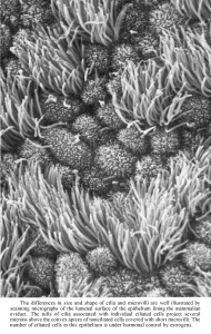
Cilia (sing. cilium) are hair-like projections on the cell surface. Their purpose is to move material across the surface of the cell. They are usually found on the apical or luminal surface of cells that line some macroscopic tubes in the body (such as the trachea or reproductive ducts).
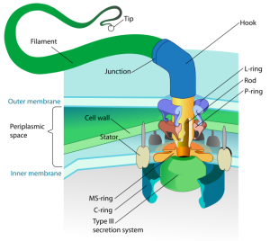
Flagella
Flagella (sing. flagellum) are whip-like projections on the cell surface. They are used to move the cell around in its environment. The only human cell that has a need to move independently is the sperm cell. Flagella are commonly found on pathogenic organisms, as seen here: Clostridium difficile, Salmonella typhi, and Giardia lamblia are shown, left to right.
Movement in Cilia & Flagella
Both cilia and flagella have the same basic structure; both are constructed of groups of microtubules running the length of the organelle in a regular pattern. At the base of both cilia and flagella, where their roots are embedded in the cell, a cross section shows a circular arrangement of microtubules in groups of three (nine triplets) at the microtubule-organizing center, the centriole.
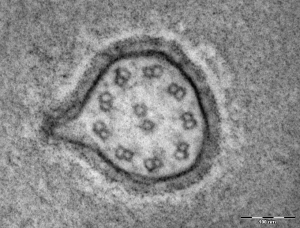
In the body of both cilia and flagella, the structure seen in cross-section changes to nine doublets (figure “8” appearance) with a pair of singlet microtubules (letter “o” appearance) in the center. Proteins such as dynein or kinesin (shown in yellow) and nexin links (red) are microtubule-associated proteins that regulate the movement of doublets past one another to create characteristic movements.
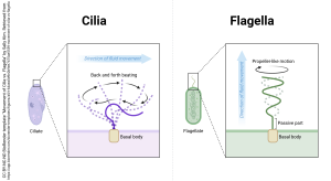
Though it’s difficult to see the difference in these proteins in diagrams or electron micrographs of cilia and flagella, molecular-level structural differences create different types of movement in the cilium vs flagellum. The base of a flagellum stays put and the ends whip around to create motion.
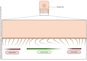
On the other hand, a cilium has a characteristic rowing motion with a power stroke and a recovery stroke. This is seen in the movie which you can access through the Canvas course. That animation, by Leanne Triezenburg, shows the molecular basis for the motion of cilia much better than these words can.
Mucus and dissolved particles in the respiratory system form a fluid layer which is moved by the respiratory cilia working together in a mucociliary escalator (also called a mucociliary apparatus). The synchronous beating of the cilia, which resembles amber waves of grain, is called a metachronal wave. This is the only mechanism to bring crap out of your lungs; if it fails, your lungs fail. We will study this in Unit 17.
Media Attributions
- U05-040 Movement of Cilia © Barnett, Cierra Memphis is licensed under a CC BY-NC-ND (Attribution NonCommercial NoDerivatives) license
- Flagellum_base_diagram-en.svg © LadyofHats is licensed under a Public Domain license
- U05-041 fawcett fig 316 p581 comparison of cilia and flagella © Fawcett, Don is licensed under a CC BY-NC-ND (Attribution NonCommercial NoDerivatives) license
- U05-044 Flagellum_of_Chara_sp_TEM_140000x © and3k and caper437 is licensed under a CC BY-SA (Attribution ShareAlike) license
- U05-046 Movement of Cilia vs. Flagella © Kim, Sally is licensed under a CC BY-NC-ND (Attribution NonCommercial NoDerivatives) license
