Membranes
Objective 7.11
7.11.1 Compare and contrast epithelial and connective tissue membranes.
7.11.2 Describe the different types of epithelial membranes: mucous, serous, and cutaneous.
7.11.3 Describe a synovial membrane.
There are two types of tissue membranes: epithelial and connective tissue.
Epithelial membranes:
- Line internal passages (mucous membranes) that lead to the exterior of the body (for example the gastrointestinal tract),
- the organs (serous membranes), and
- epithelial tissue membranes are linings that cover the outside of the body (the cutaneous membrane, i.e. skin),
Connective tissue membranes encapsulate an organ (such as the kidney) or line freely moveable joints (synovial membranes).
Epithelial membranes are flat and flexible sheets, which form a lining where parts of the body come together. They consist of epithelium plus underlying areolar connective tissue.
Mucous membranes line the interface between the body cavities and the outside world, which include the digestive, respiratory, excretory, and reproductive tracts. Mucus membranes consist of epithelial cells attached to a basement membrane + connective tissue deep to the epithelium. The connective tissue layer of mucous membranes is called a lamina propria, which literally means “proper thin sheet.” This layer helps support the fragile epithelial tissue. Mucus is produced by epithelial exocrine glands which covers the epithelial layer, hence the name “mucous membrane.”
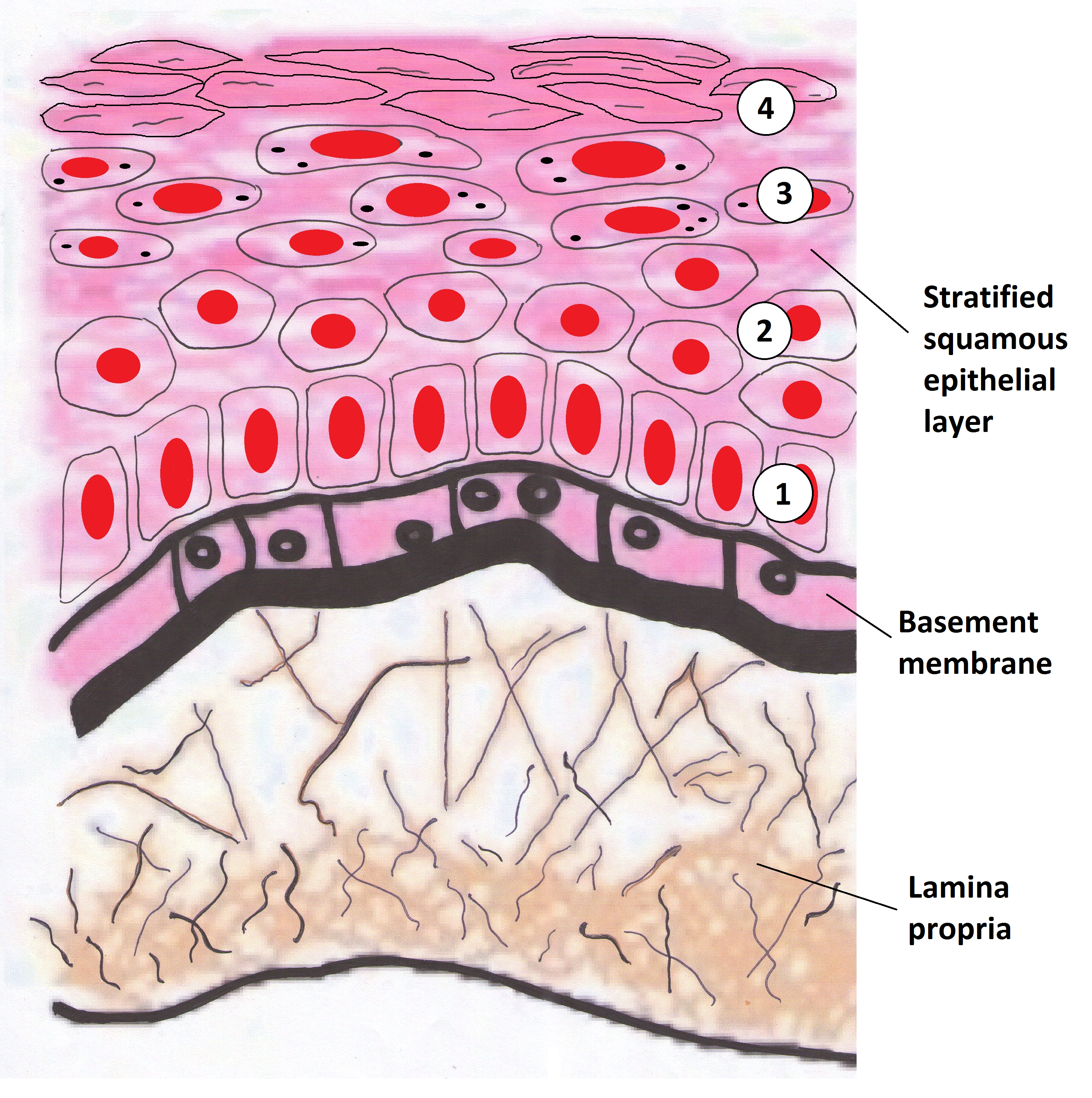
In this depiction of the oral mucosa we see layers of stratified epithelium attached to a basement membrane. Deep to that is connective tissue, the lamina propria.
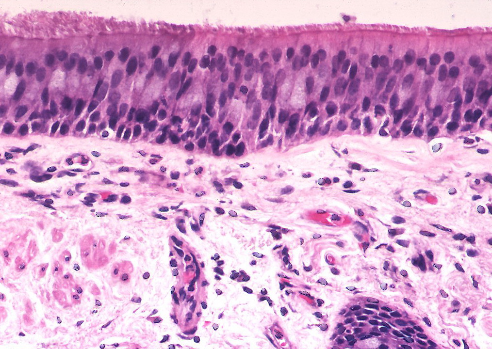
The respiratory mucosa, as seen in this light micrograph, consists of pseudostratified columnar epithelium with the lamina propria deep to the epithelial cells. If you look closely, you can see the basement membrane.
Serous membranes form a lining between the body wall and internal organs. They consist of a single layer of cells called the mesothelium, which is attached to a supporting layer of connective tissue.
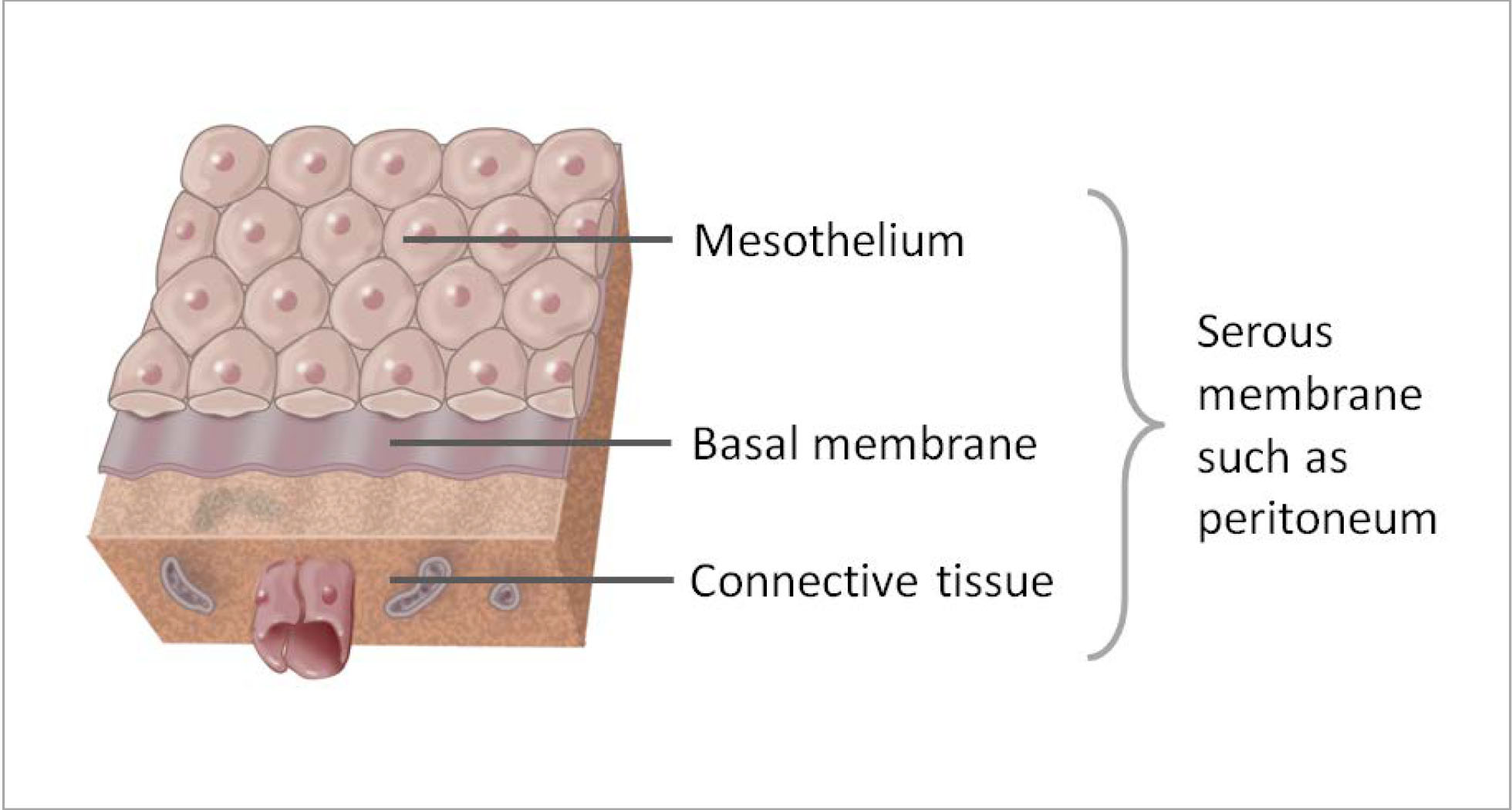
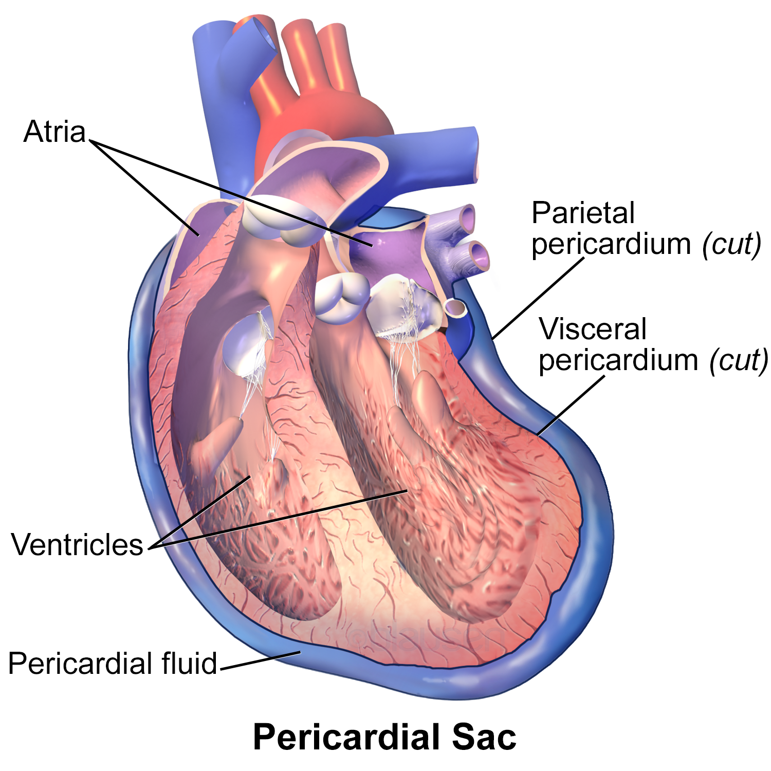
These membranes literally form a bag. The inner layer of the bag, the visceral layer, lies directly on the organ. The outer layer of the bag, the parietal layer, lies away from the organ. The mesothelial cells secrete serous fluid, which lies between the two layers. This fluid allows movement and lubrication.
Specific names are given to serous membranes. Those that surround the lungs are called the pleural membranes, surrounding the heart, pericardial membranes, and surrounding the GI tract, the peritoneal membrane.
The cutaneous membrane, or skin, covers the outside of the body away from cavities. The epithelial layer is composed of stratified squamous epithelium. Deep to this epithelium is connective tissue.

In the below image, we see the epithelial layer of the skin, the epidermis. Deep to that is the connective tissue, the dermis.
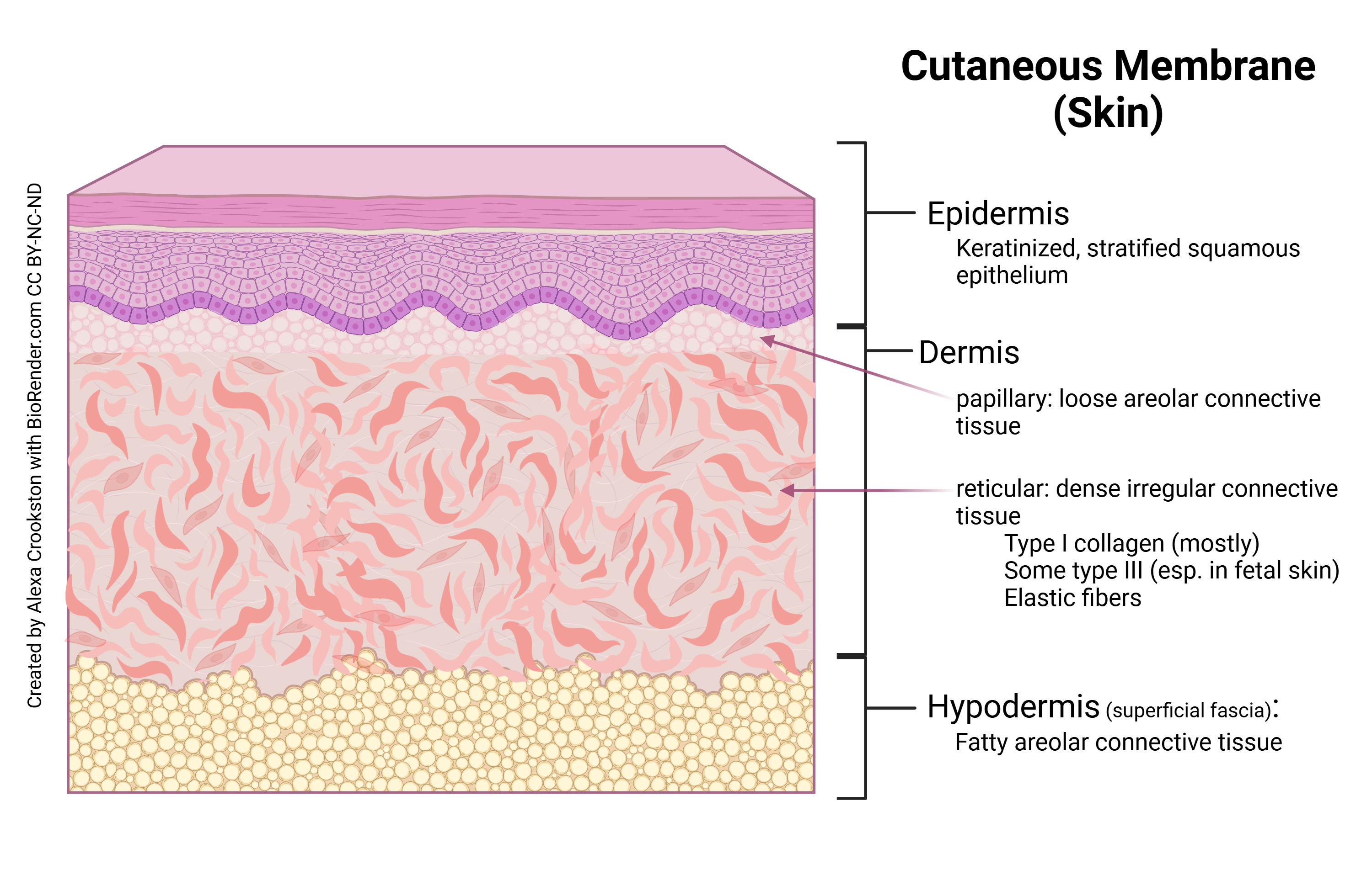
We will be studying the skin in depth in the next unit, the Integumentary System.
Synovial membranes line the space between two bones forming a joint. Joints allow movement while preventing the bones from rubbing against one another and causing damage to the bone.
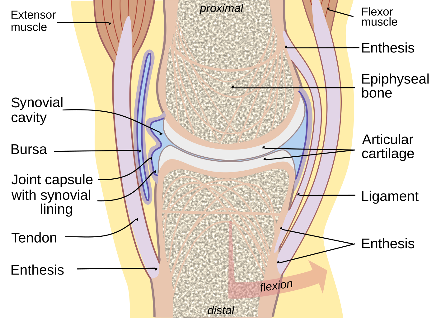
Synovial membranes lack an epithelial layer and are made up only of connective tissue. The connective tissue contains specialized cells called synoviocytes. These cells secrete synovial fluid that lubricates the joint.
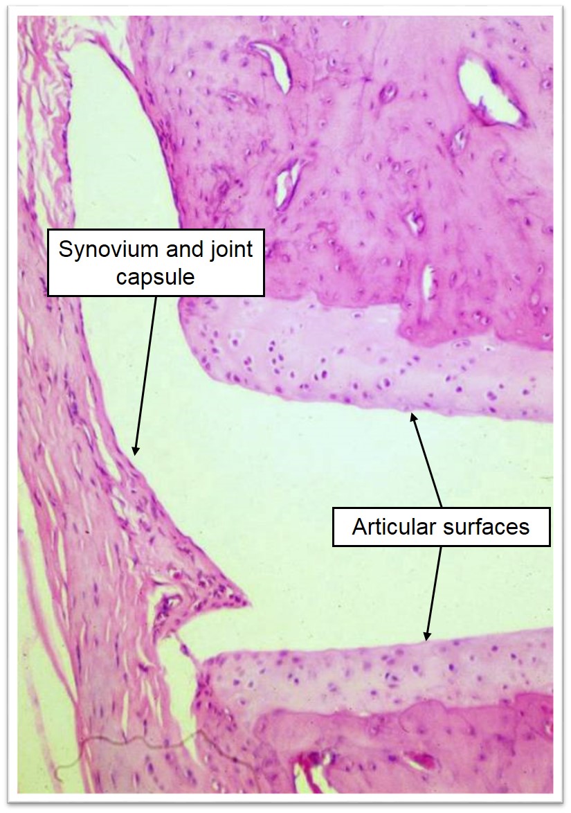
In this histological section of a synovial joint, the synovial joint cavity, capsule and synoviocytes (synovium) can be visualized.

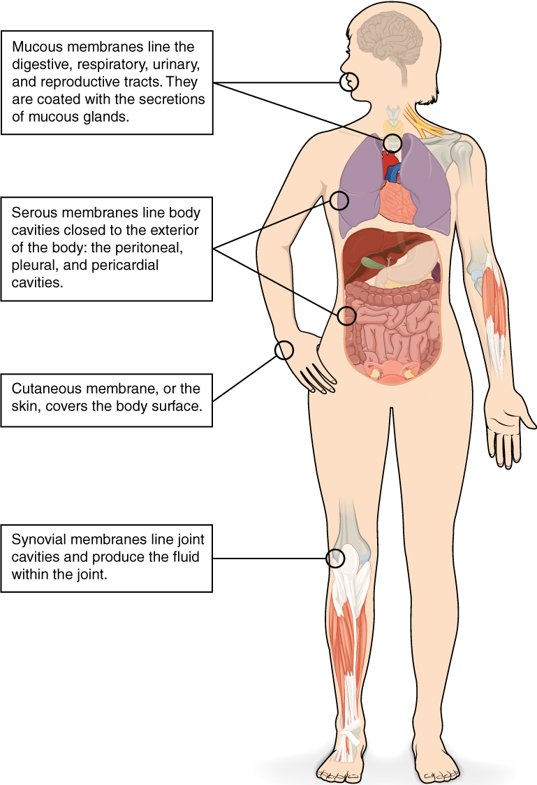
This image summarizes the types of membranes we’ve been discussing and their locations.
Media Attributions
- U07-073 Oral_mucosa © Wiki-Minor is licensed under a CC BY-SA (Attribution ShareAlike) license
- U07-074 Respiratory mucosa © Rosen, Yale is licensed under a CC BY-SA (Attribution ShareAlike) license
- U07-075 Histology of serous membranes-1 © O.P. Paul Gobee is licensed under a CC BY-NC-SA (Attribution NonCommercial ShareAlike) license
- U07-076 Blausen_0724_PericardialSac © Blausen Medical Communications, Inc. is licensed under a CC BY (Attribution) license
- Screenshot 2025-03-20 at 9.22.43 AM (2) © Betts, J. Gordon; Young, Kelly A.; Wise, James A.; Johnson, Eddie; Poe, Brandon; Kruse, Dean H. Korol, Oksana; Johnson, Jody E.; Womble, Mark & DeSaix, Peter is licensed under a CC BY (Attribution) license
- U07-078 Cutaneous Membrane (1) © Crookston, Alexa is licensed under a CC BY-NC-ND (Attribution NonCommercial NoDerivatives) license
- U07-079 Joint © MadHero88 is licensed under a CC BY-SA (Attribution ShareAlike) license
- U07-080 synovial joint histology © Jennings, Ryan and Premanandan, Christopher is licensed under a CC BY-NC (Attribution NonCommercial) license
- U07-081 48fd4d691119908e3d6b24f7ecfede7bb0fb22e4 2 © OpenStax is licensed under a CC BY (Attribution) license

.jpg)