Membrane Proteins
Objective 4.4
4.4.1 Name classes of membrane proteins and describe their function.
Now, let’s turn to the important role played by proteins in the properties of the cell membrane. We used the terms peripheral and integral to indicate the microscopic anatomy of the proteins. Now we’re going to look at the functional (physiological) role of membrane proteins.
We’ve made brief reference to some of these, but now we’re going to look at categories of membrane proteins: 1) ion channels; 2) receptors; 3) carriers; 4) enzymes; 5) linkers; and 6) cell identity markers. Note the overlap between the anatomical description (integral or peripheral) and the physiological category in each case.
Ion Channels
Charged ions (Na+, K+, Cl–, H+, Ca++, HCO3–, and many others) cannot cross the lipid bilayer without assistance. In order to cross the cell membrane, they need ion channels, proteins that span the lipid bilayer and allow these ions to pass. In general, ion channels are specific for one or two types of ions (for example, there are Na+ channels that do not easily permit K+ to pass). Another interesting feature of some of these channels is that they’re gated: they open or close on demand.
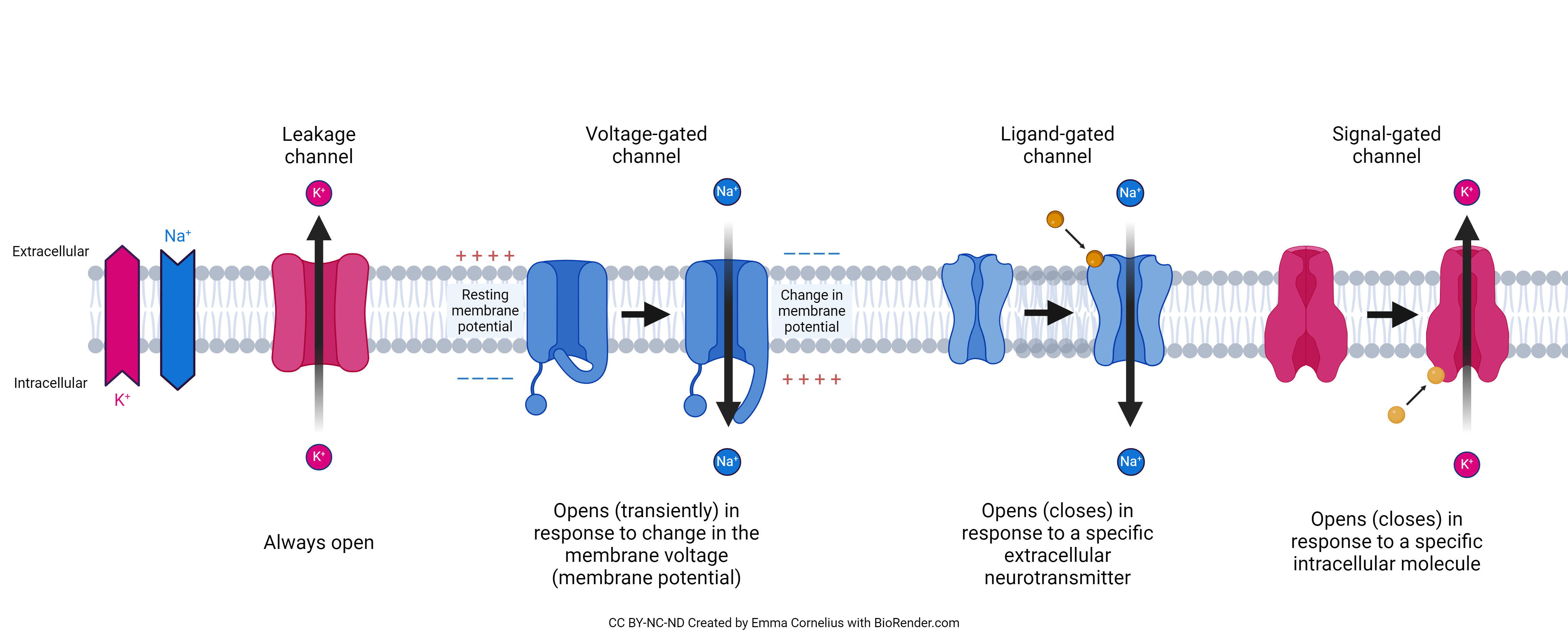
This picture shows different types of ion channels that are used by neurons as they receive information. Ligand-gated channels need a chemical (called a ligand) to open them. These are also called receptor proteins because they are picking up a signal from outside of the cell. Ion channels open or close depending on the signal. We’ll discuss receptor proteins in a little more detail coming up. Mechanically-gated channels are opened by pressure on the surface of the skin or in the inner ear. Leak channels are always open and allow ions to leak into the cell at a steady rate, which maintains a charge that nerve cell function depends on. Voltage-gated channels open by a change in the distribution of electrically charged ions between the inside and outside of a cell. These are also important parts of the cell membrane of neurons, used to send information over long distances.
Receptor Proteins
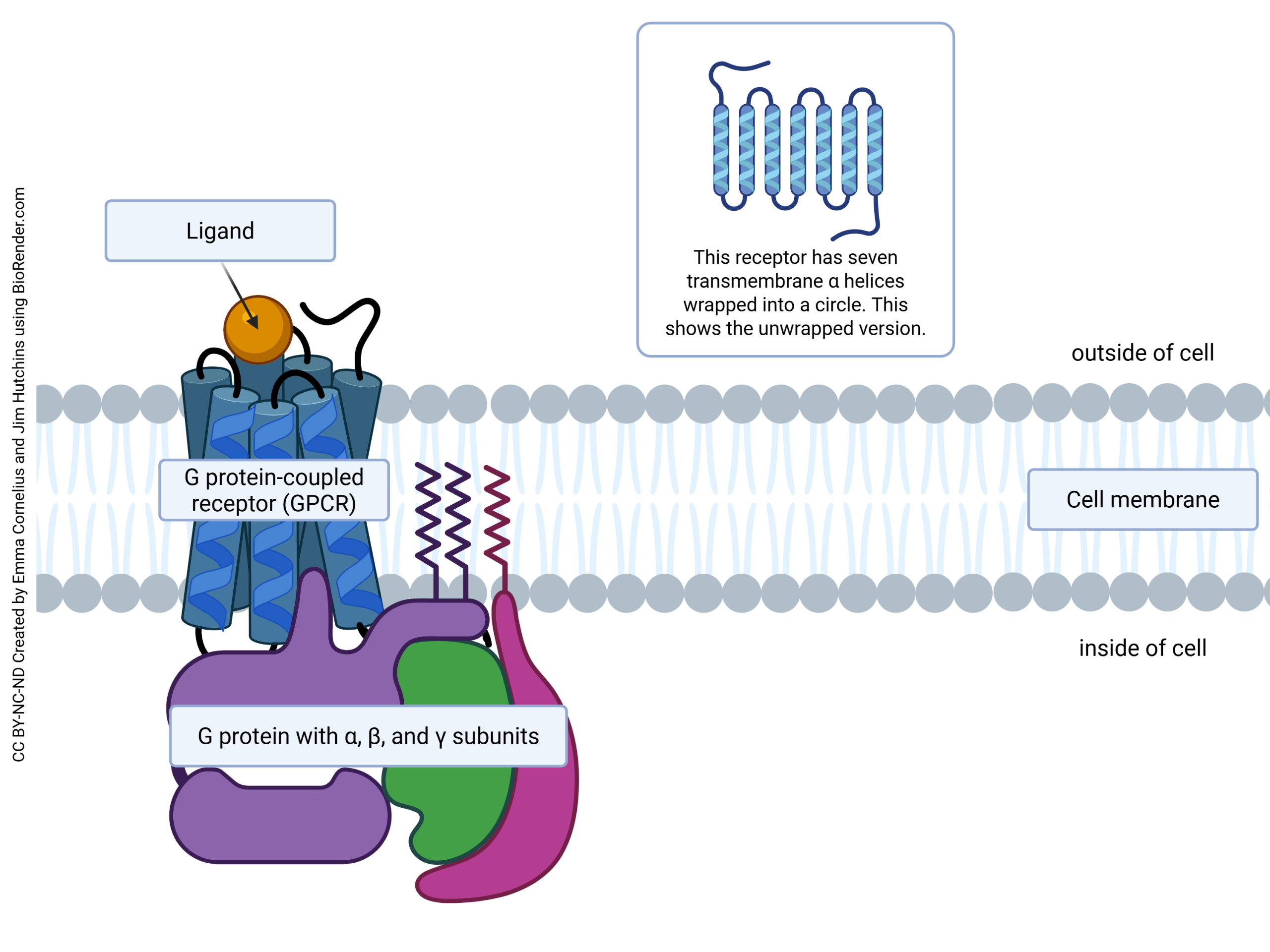
Receptor proteins receive a signal (ligand) which will make a change within the cell. We mentioned receptor proteins above that may open or close an ion channel. Another type of receptor is a G protein-coupled receptor. When this type of receptor receives a signal, the G protein bound to the receptor acts like a switch within the cell, turning on or off certain cellular processes. We’ll discuss receptors in more depth in the nervous and endocrine units.
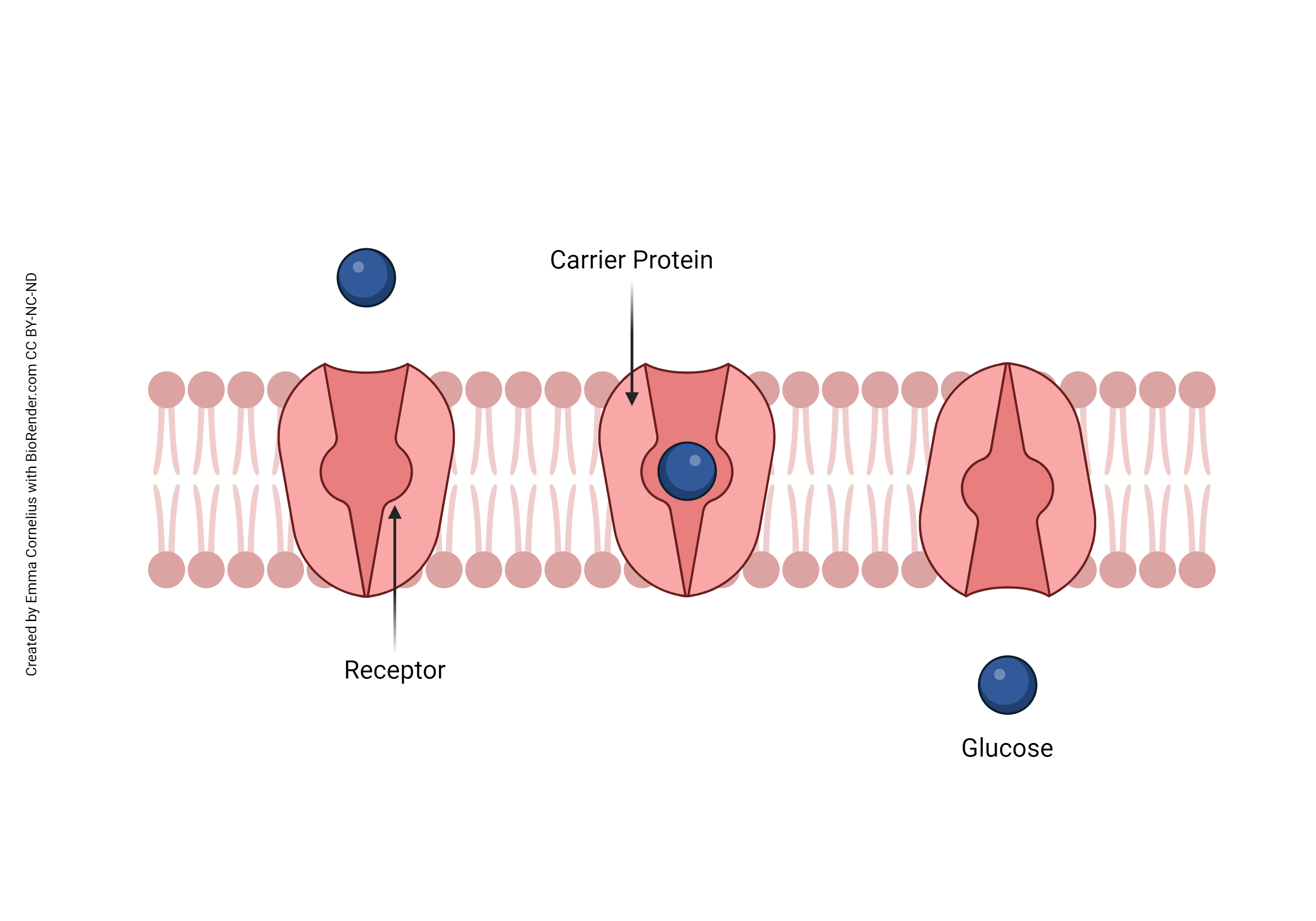
Carrier Proteins
Some membrane proteins are carriers. Glucose is a charged molecule and cannot easily pass through the charged polar heads of the cell membrane. Instead, glucose is carried across the lipid bilayer. There are two possibilities: either glucose is carried down its concentration gradient, which does not require energy input, or glucose is carried up its concentration gradient, which requires energy. If energy is required, it can be obtained either from the energy currency (ATP) or from the movement of another ion down its concentration gradient. We’ll discuss these examples more in objectives 4 and 5.
Enzymes
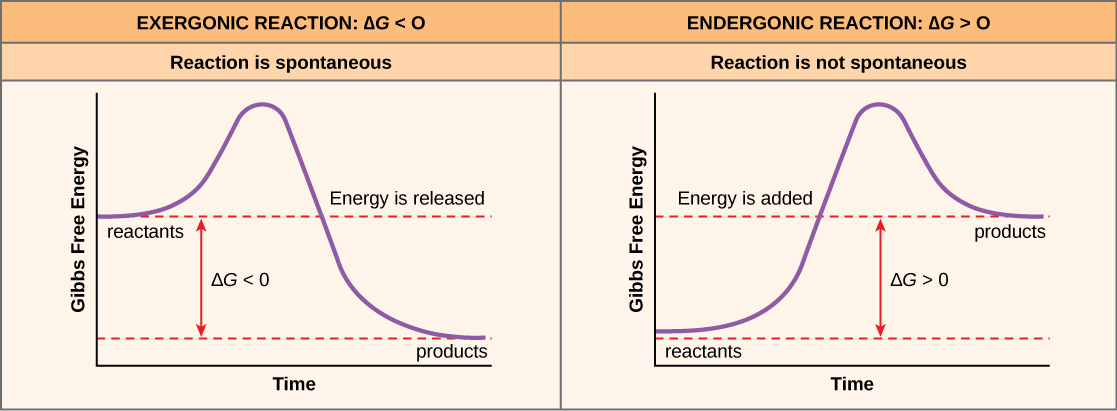
We looked at proteins called enzymes in the last unit. Remember that enzymes catalyze chemical reactions; they can’t and don’t change the energy profile of the reaction, but instead lower the activation energy (the “bump” in the energy curve).
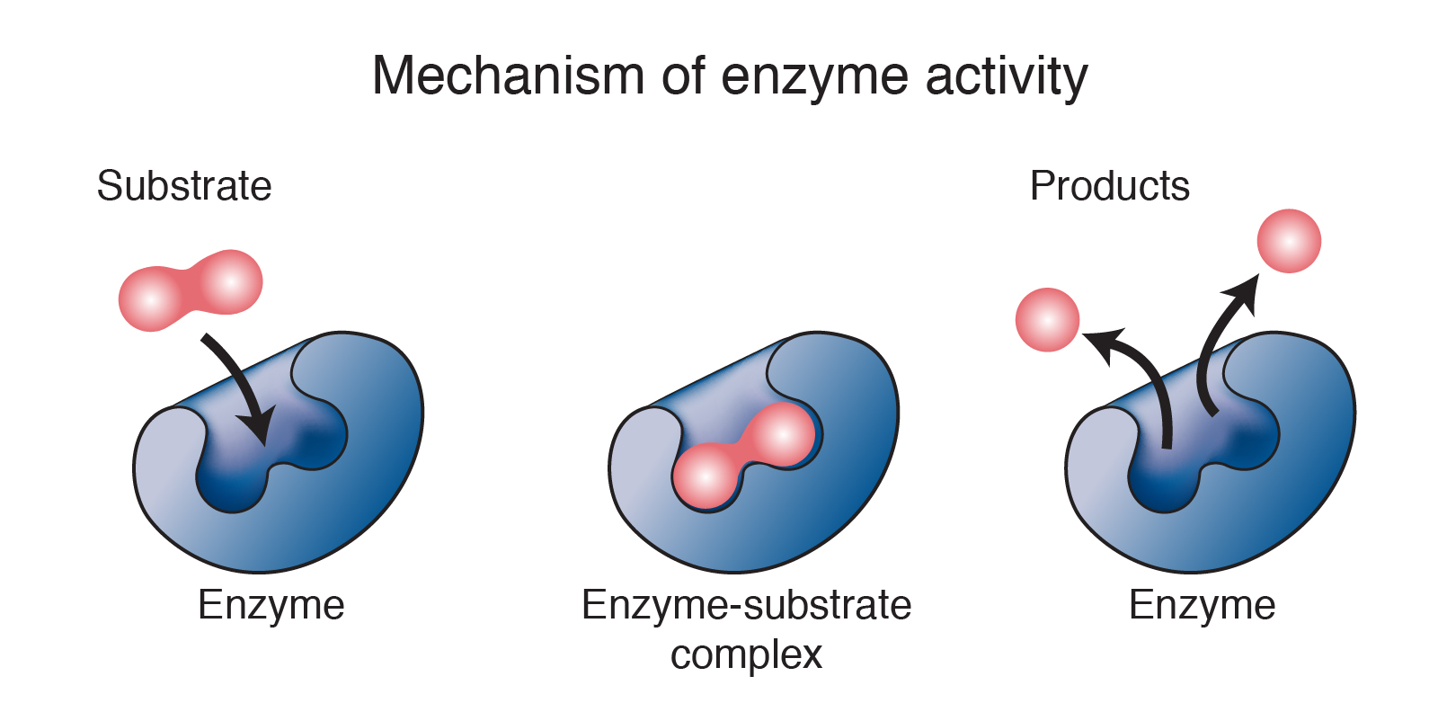
Some of these enzymes are found on the cell surface, where they carry out necessary chemical reactions. For example, in the intestine, where milk sugar (lactose) is broken down, the enzyme responsible for the breakdown (lactase) is found on the surface of intestinal cells.
Linker Proteins
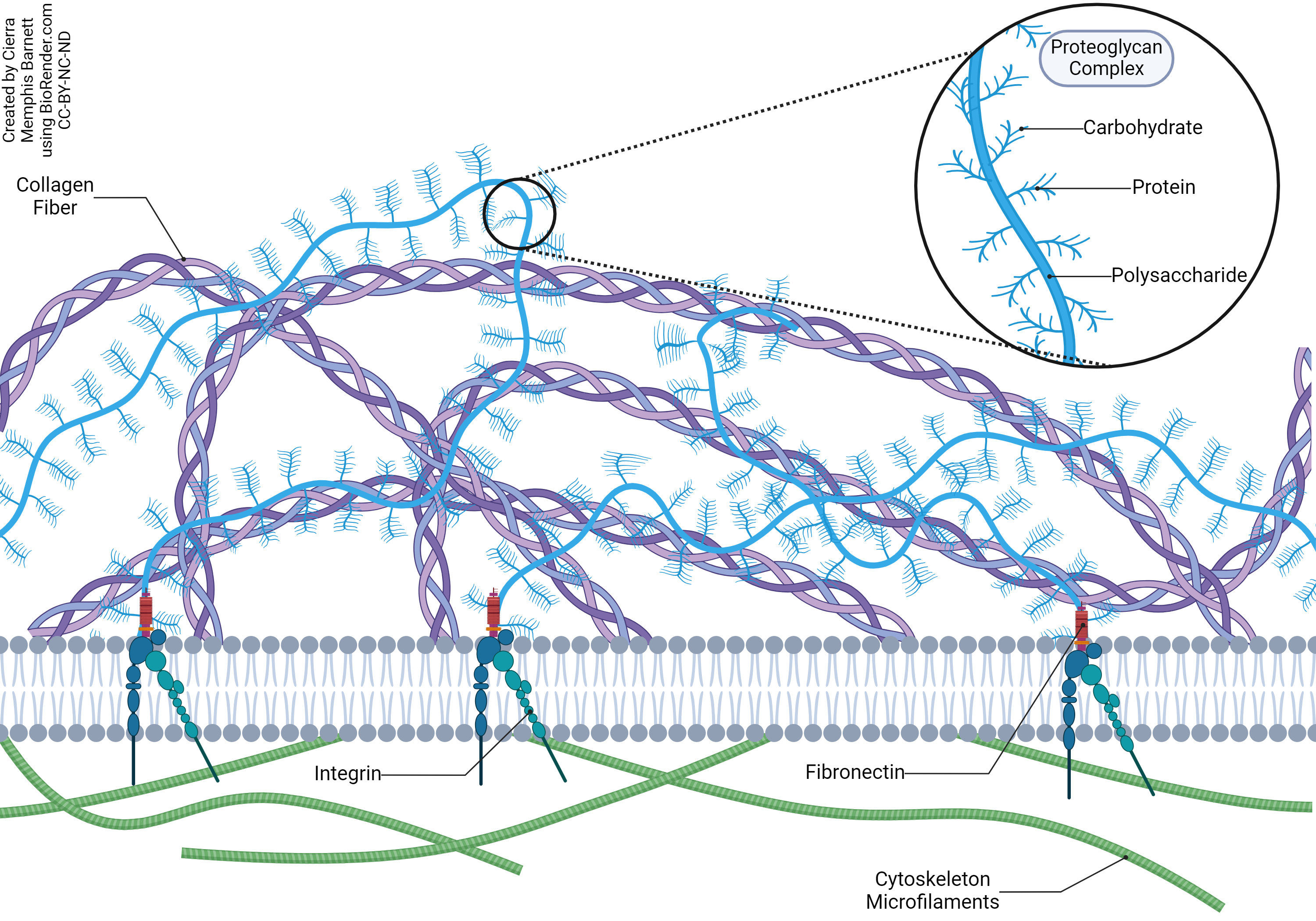
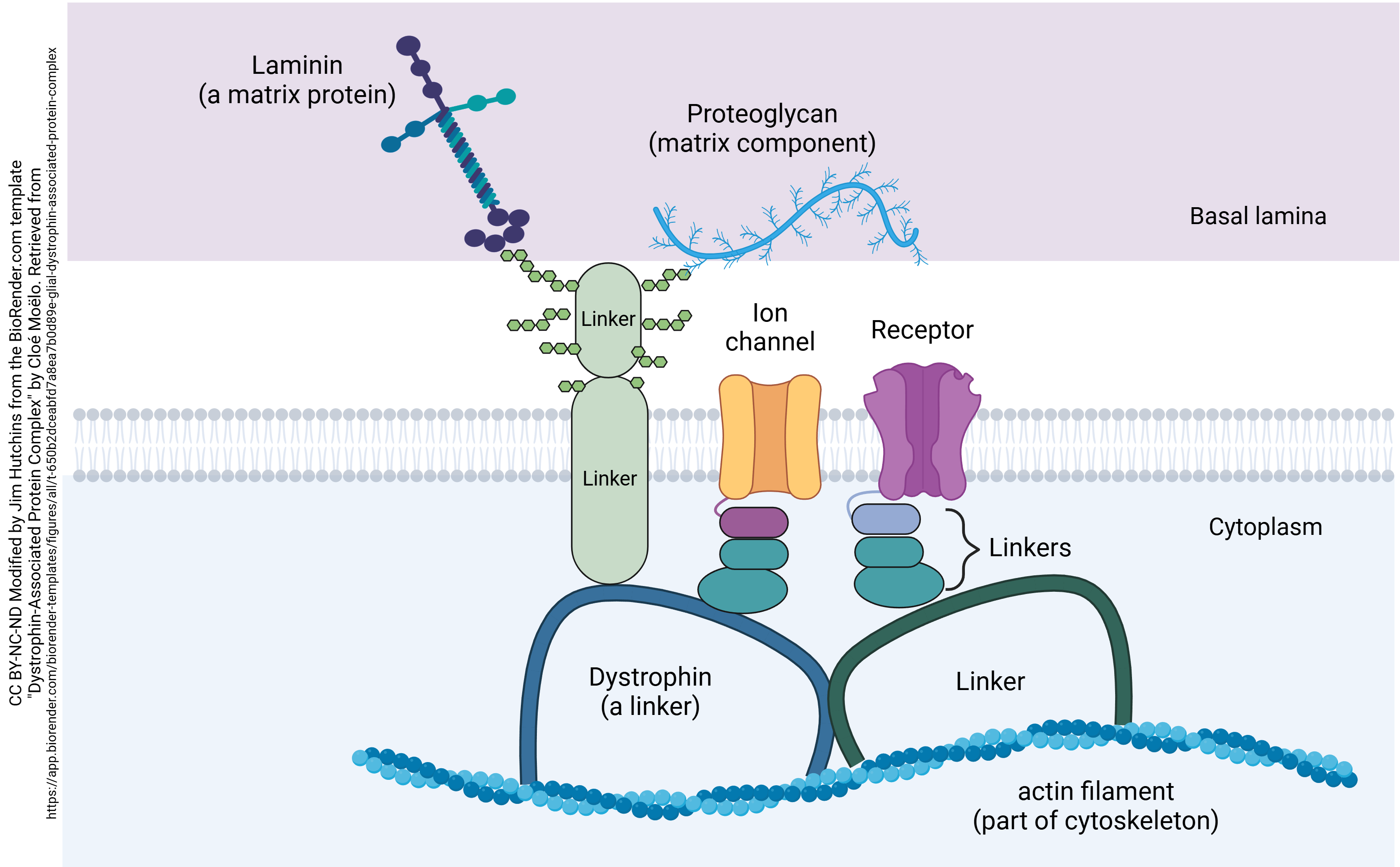
Cells that are arranged into tissues and organs must bind themselves together. A combination of integral and peripheral proteins are responsible for this linking function. On the intracellular side, linkers join integral membrane proteins to the cytoskeleton, the structural proteins within the cell. On the extracellular side, linkers join the same integral membrane proteins to the connective tissue matrix or to other cells.
For example, integrin is a linker protein that facilitates cell-extracellular matrix adhesions.
Fibronectin, also shown here, is a linker protein which binds to integrin and joins the collagen fibers of the extracellular matrix (connective tissue) to integrin, and then integrin is bound to the cytoskeleton, linking the strong fiber networks of the outside of the cell to the strong fiber networks inside the cell. Proteins such as dystrophin, which is involved in the disease muscular dystrophy, bind transmembrane proteins to the cytoskeleton.
Cell Marker Proteins
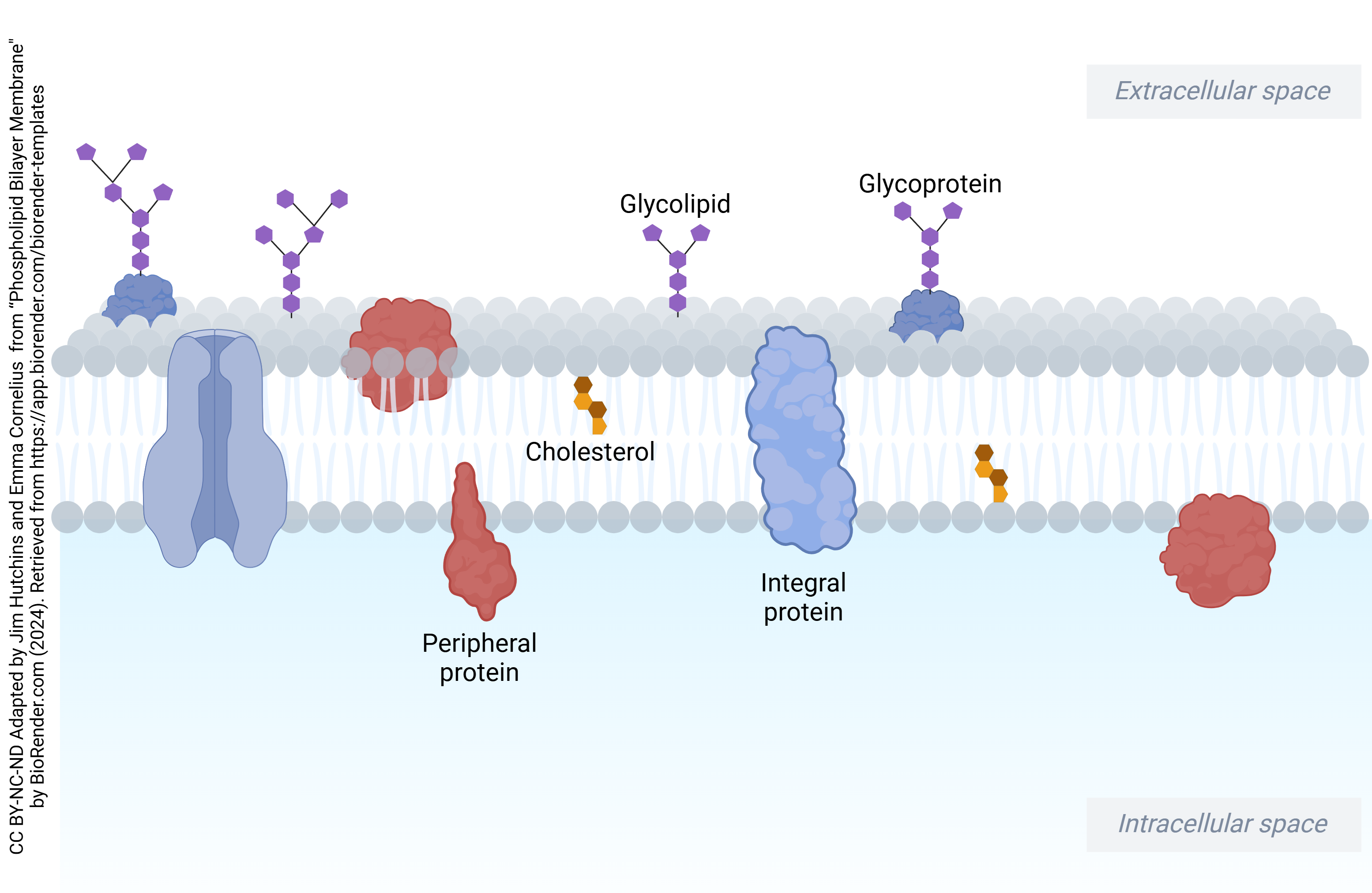
In this image of the cell membrane, glycoproteins are used to identify the cells. Cells need to be able to identify themselves in two ways: like baggage tags, they need to identify the owner, and they need to have an origin address or a destination.
This function is achieved by cell marker proteins. Most cell markers consist of a complex, branched sugar polymer attached to a transmembrane protein. This combination is called a glycoprotein (glyco-: “sugar’). If you pick up the wrong bag at the airport, a security guard will stop you. If a cell ends up trying to occupy the wrong organ, or a foreign cell is deliberately or mistakenly transplanted, the security guards of the body — the immune cells — will stop the invasion.
Media Attributions
- U04-023 same as U13-005 Ion Channels © Cornelius, Emma is licensed under a CC BY-NC-ND (Attribution NonCommercial NoDerivatives) license
- U04-024 GPC Receptor Proteins © Cornelius, Emma and Hutchins, Jim is licensed under a CC BY-NC-ND (Attribution NonCommercial NoDerivatives) license
- U04-025 carrier proteins © Cornelius, Emma is licensed under a CC BY-NC-ND (Attribution NonCommercial NoDerivatives) license
- U04-026 enzyme © National Human Genome Research Institute is licensed under a Public Domain license
- U04-027 Exergonic_endergonic reactions © Thermodynamics Forum is licensed under a CC BY-NC-ND (Attribution NonCommercial NoDerivatives) license
- U04-028 Linker Proteins © Barnett, Cierra Memphis is licensed under a CC BY-NC-ND (Attribution NonCommercial NoDerivatives) license
- U04-028a Linker proteins © Hutchins, Jim & Moelo, Cloe is licensed under a CC BY-NC-ND (Attribution NonCommercial NoDerivatives) license
- U04-012 and 029 phospholipid bilayer © Hutchins, Jim & Cornelius, Emma is licensed under a CC BY-NC-ND (Attribution NonCommercial NoDerivatives) license
