Lower Extremity Muscles
Objective 10.11
10.11.1 Give the origin, insertion, and action of the major muscles of the lower extremity.
10.11.2 Identify these muscles on a picture or image.
Lower Extremity
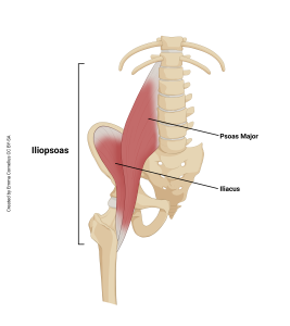
Iliopsoas
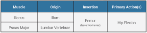
The iliopsoas muscle is actually two muscles: iliacus and psoas major. Most people refer to this muscle as their ‘hip flexor’. If you were a pig, we’d call this your “tenderloin”. They have different origins but come together to attach on the lesser trochanter of the femur.
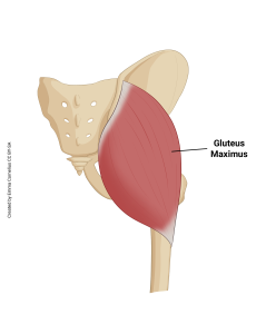
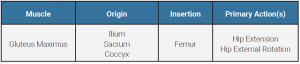
Gluteus Maximus
The gluteus maximus is one of the largest and strongest muscles in the body. This muscle is a good example of being named for its location and size. This is also a common site for intramuscular (IM) injections, especially for larger volumes that dissipate over a longer period (such as penicillin or gamma globulin).
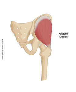
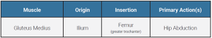
Gluteus Medius
The gluteus medius is an important muscle for hip and knee stability and control. It is located on the lateral hip. Much like the deltoid, it is a common location for injections.
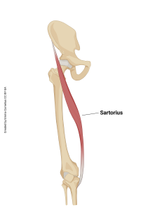
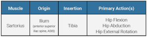
Sartorius
The sartorius is the longest muscle in the body.
Muscle Compartments
In studying the muscles of the thigh, it is helpful to know that the thigh is divided into three compartments. Blood vessels and nerves run in “seams” between the compartments.
- Medial compartment contains adductors (purple in image)
- Anterior compartment contains quadriceps (blue in image)
- Posterior compartment contains hamstrings (green in image)
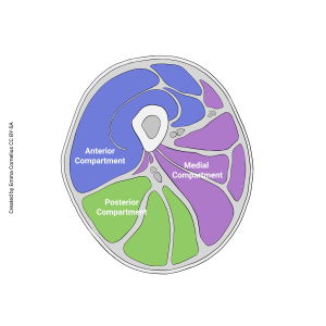
Other limbs, such as the lower leg, have similar compartments but we will not study them in this course.
Clinical Connection
Compartment Syndrome
If pressure builds up in one of these compartments due to injury it can cause serious damage by cutting off nerve and blood supply. If pressure becomes severe, a surgery called a fasciotomy is performed which is fileting through the compartments to release pressure.
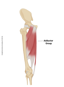
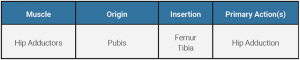
Hip Adductor Group
The adductors are comprised of the muscles in your medial thigh. They are housed in the medial compartment. Much like the wrist flexors and extensors, we are grouping the hip adductors together. These are typically referred to as your “groin muscles”.
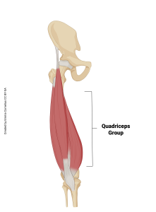
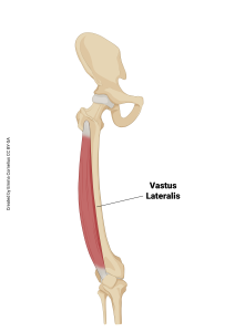
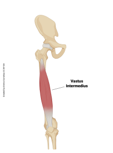
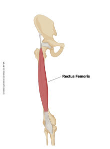
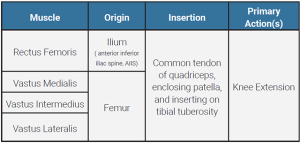
Quadriceps Group
The quadriceps muscle group is comprised of the anterior thigh muscles. These muscles are housed in the anterior compartment of your thigh.

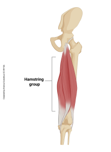
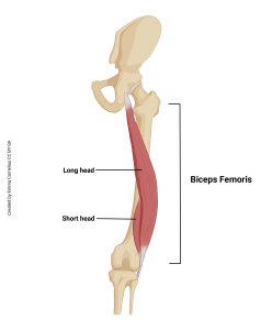
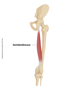
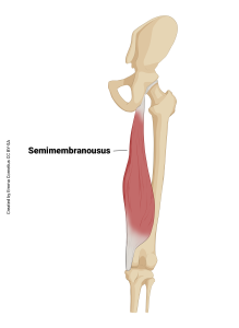
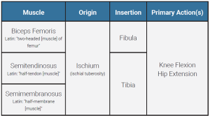
Hamstring Group
The hamstring muscle group is comprised of the posterior thigh muscles. These muscles are housed in the posterior compartment of your thigh. They have unique names, which are explained in the table below.
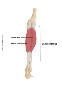
Gastrocnemius
The gastrocnemius is the muscle that is often referred to when talking about the “calf muscles”.

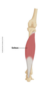
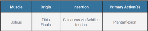
Soleus
The soleus is deep to the gastrocnemius. Much like the brachialis, the soleus does not get much credit for the work it does because you do not see much of it from the surface of the body.
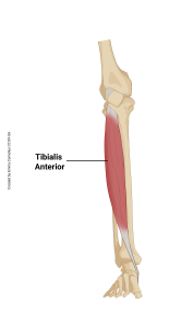
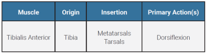
Tibialis Anterior
The tibialis anterior is the muscle on the anterior leg (crus). This muscle performs dorsiflexion which is important for walking so you don’t trip over your feet. This muscle is often impaired as part of a stroke and patients develop drop foot.
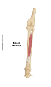
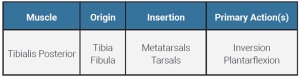
Tibialis Posterior
The tibialis posterior is the shin-splints muscle. It helps support your arch and when you run or jump a lot, it can get overworked and painful.
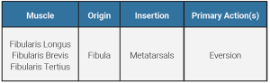
Fibularis/Peroneal Group
The fibularis muscle group consists of three muscles. These muscles are also called the peroneal group. You can use the names interchangeably. Here,we will use fibularis to name the muscles based on their anatomic location. These muscles work together to prevent your ankle from rolling inward (inversion), the most common type of ankle sprain.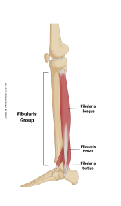
Media Attributions
- U10-060 Iliopsoas © Cornelius, Emma is licensed under a CC BY-SA (Attribution ShareAlike) license
- U10-061 ileopsoas origin and insertion table © Bizzell, Lizz is licensed under a CC BY-SA (Attribution ShareAlike) license
- U10-062 gluteus maximus © Cornelius, Emma is licensed under a CC BY-SA (Attribution ShareAlike) license
- U10-063 gluteus maximus origin and insertion table © Bizzell, Lizz is licensed under a CC BY-SA (Attribution ShareAlike) license
- U10-064 gluteus medius © Cornelius, Emma is licensed under a CC BY-SA (Attribution ShareAlike) license
- U10-065 gluteus medius origin and insertion table © Bizzell, Lizz is licensed under a CC BY-SA (Attribution ShareAlike) license
- U10-066 Sartorius © Cornelius, Emma is licensed under a CC BY-SA (Attribution ShareAlike) license
- U10-067 sartorius origin and insertion table © Bizzell, Lizz is licensed under a CC BY-SA (Attribution ShareAlike) license
- U10-068 thigh cross section © Cornelius, Emma is licensed under a CC BY-SA (Attribution ShareAlike) license
- U10-069 Adductor Group © Cornelius, Emma is licensed under a CC BY-SA (Attribution ShareAlike) license
- U10-070 hip adductors origin and insertion table © Bizzell, Lizz is licensed under a CC BY-SA (Attribution ShareAlike) license
- U10-071 Quadriceps Group © Cornelius, Emma is licensed under a CC BY-SA (Attribution ShareAlike) license
- U10-073 Vastus Lateralis © Cornelius, Emma is licensed under a CC BY-SA (Attribution ShareAlike) license
- U10-075 Vastus Intermedius © Cornelius, Emma is licensed under a CC BY-SA (Attribution ShareAlike) license
- U10-074 Vastus Medialis © Cornelius, Emma is licensed under a CC BY-SA (Attribution ShareAlike) license
- U10-007 Rectus Femoris © Cornelius, Emma is licensed under a CC BY-SA (Attribution ShareAlike) license
- U10-072 quadriceps group origin and insertion table © Bizzell, Lizz is licensed under a CC BY-SA (Attribution ShareAlike) license
- U10-076 Hamstring group © Cornelius, Emma is licensed under a CC BY-SA (Attribution ShareAlike) license
- U10-078 biceps femoris © Cornelius, Emma is licensed under a CC BY-SA (Attribution ShareAlike) license
- U10-079 Semitendinosus © Cornelius, Emma is licensed under a CC BY-SA (Attribution ShareAlike) license
- U10-080 semimembranosus © Cornelius, Emma is licensed under a CC BY-SA (Attribution ShareAlike) license
- U10-077 hamstring group origin and insertion table © Bizzell, Lizz is licensed under a CC BY-SA (Attribution ShareAlike) license
- U10-081 Gastrocnemius © Cornelius, Emma is licensed under a CC BY-SA (Attribution ShareAlike) license
- U10-082 gastroc origin and insertion table © Bizzell, Lizz is licensed under a CC BY-SA (Attribution ShareAlike) license
- U10-083 soleus © Cornelius, Emma is licensed under a CC BY-SA (Attribution ShareAlike) license
- U10-084 soleus origin and insertion table © Bizzell, Lizz is licensed under a CC BY-SA (Attribution ShareAlike) license
- U10-085 tibialis anterior © Cornelius, Emma is licensed under a CC BY-SA (Attribution ShareAlike) license
- U10-086 tibialis ant origin and insertion table © Bizzell, Lizz is licensed under a CC BY-SA (Attribution ShareAlike) license
- U10-087 tibialis posterior © Cornelius, Emma is licensed under a CC BY-SA (Attribution ShareAlike) license
- U10-088 tibialis post origin and insertion table © Bizzell, Lizz is licensed under a CC BY-SA (Attribution ShareAlike) license
- U10-089 fibularis group © Cornelius, Emma is licensed under a CC BY-SA (Attribution ShareAlike) license
- U10-090 fibularis origin and insertion table © Bizzell, Lizz is licensed under a CC BY-SA (Attribution ShareAlike) license
