Features of Epithelial Tissues
Objective 7.2
7.2.1 Describe basic structural features of epithelial tissue.
7.2.2 Describe naming conventions for the cell shape and layering of epithelial tissue.
Epithelia form different types of barriers. Each has several features in common. Epithelial cells have two surfaces:
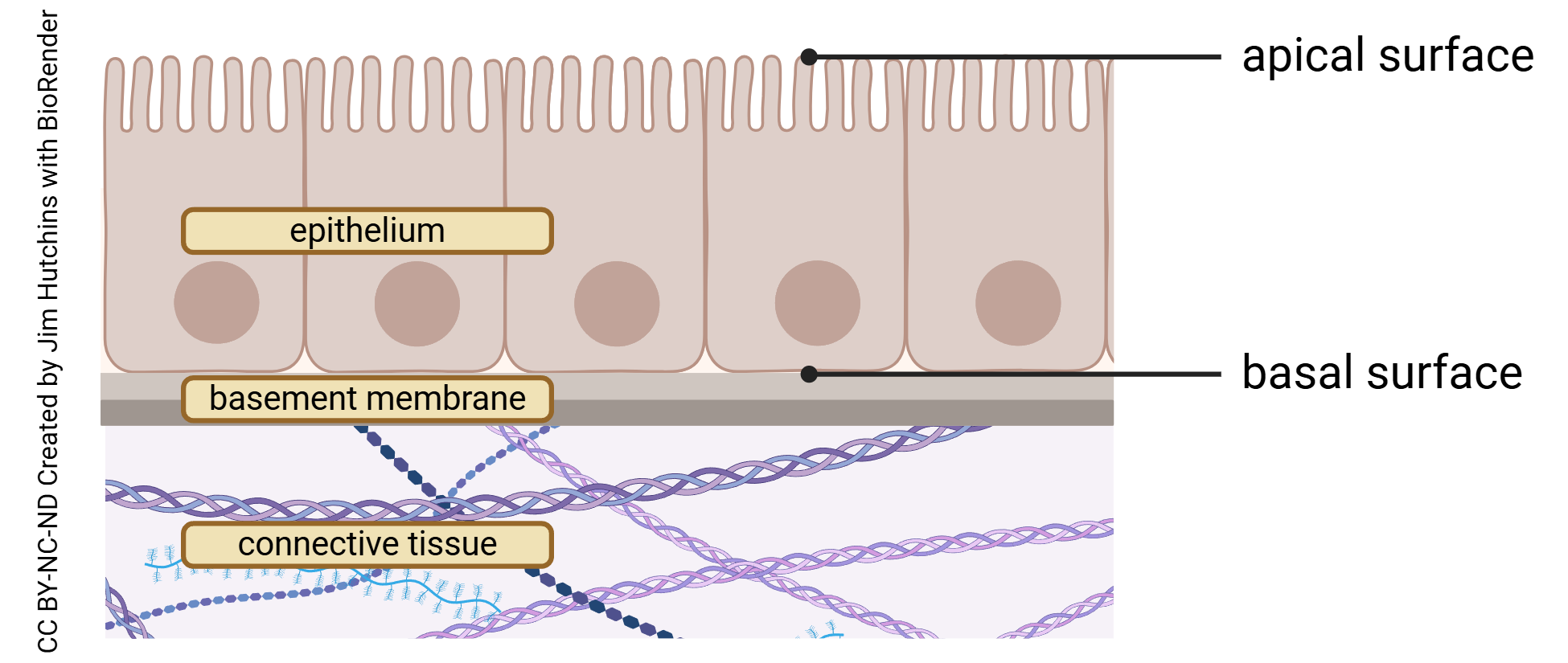
- an apical surface (Latin apex: “top, pinnacle”) that faces the outside world; and
- a basal surface (note the words “base” and “basement” here) that rests on, and is attached to, the basement membrane.
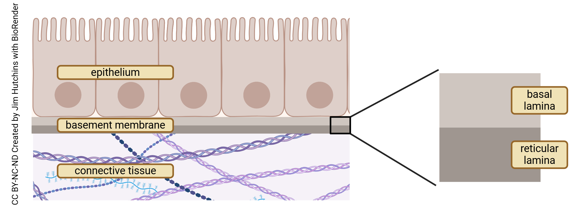
The basement membrane, in turn, has two thin layers, each called a lamina (Latin: “thin plate” or “sword blade”). The basal lamina is nearest the epithelial cells, and the reticular lamina (Latin: reticulum, “net” or “mesh”) is a bit deeper.
basement membrane = basal lamina + reticular lamina
Beneath the basement membrane is a layer of connective tissue. Blood vessels and nerves are also found in the connective tissue layer.
In some epithelial tissue, cells are layered. Cells beneath those on the apical surface may appear to have a slightly different shape, so we just look at the cell types on the apical (uppermost) surface (those that face the outside world or the lumen). A key feature is whether all cells are in contact with the basement membrane. 
The following terms describe the arrangement of the cells:
- simple: the cells are a single layer, and all are in contact with the basement membrane;
- pseudostratified: (Greek ψευδης, pseudis–: “false, lying, untrue”) even though the cells appear to have layers, each cell is in contact with the basement membrane.
- stratified: (Latin stratum: “layer”) the cells are in layers, so only the lowest layer is in contact with the basement membrane.
Epithelial tissues are described by the arrangement and shape of the cells at the apical surface.
The following terms describe the shape of the cells. If there are multiple layers (i.e., the epithelium is stratified), then the shape of the outermost (apical) cells are used to give the name.
- squamous (Latin squamosus: “scale”, “fish scale”) cells, as the name suggests, are flat and shaped like fish scales. They are wider than they are tall.
- cuboidal cells are as wide as they are tall, like little ice cubes.
- columnar cells, like a column, are taller than they are wide.
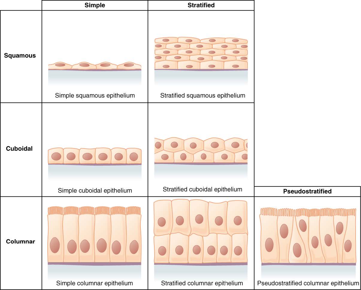
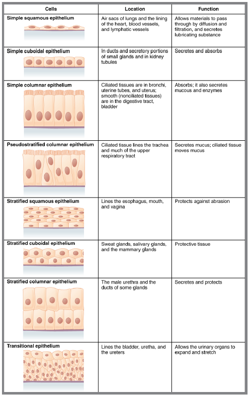
Anatomists combine the arrangement and shape of the cells to describe the different epithelial types. When we combine the three possible names for cell arrangements and the three possible names for cell shape, there are nine (3 x 3 = 9) theoretical possibilities. In reality, the only shapes of cells found in a pseudostratified epithelium are columnar cells, so there are seven possible combinations. Finally, we add back an eighth type; transitional epithelia contain cells that change shape, depending on whether the organ is enlarged or shrunken.
We will next look at each of these eight epithelial types in order, and give an example where each may be found.
Media Attributions
- U07-003 labeled generic epithelium © Hutchins, Jim is licensed under a CC BY-NC-ND (Attribution NonCommercial NoDerivatives) license
- U07-004 basement membrane parts © Hutchins, Jim is licensed under a CC BY-NC-ND (Attribution NonCommercial NoDerivatives) license
- U07-005 text box © Bizzell, Lizz is licensed under a CC BY-SA (Attribution ShareAlike) license
- U07-006 © OpenStax is licensed under a CC BY (Attribution) license
- U07-007 epithelium types table © Kathy Newton is licensed under a CC BY-SA (Attribution ShareAlike) license
